
Today
8:30 AM to 5:00 PM.
Description
Optometrists are not doctors but are trained in examining eyes and testing your vision to assess your need for glasses or contact lenses.
Opticians read prescriptions for visual correction, order lenses and dispense eye glasses and contact lenses.
Consultants
Note: Please note below that some people are not available at all locations.
-

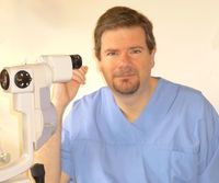
Dr David Dalziel
Ophthalmologist, Clinical Director Ophthalmology Northland District Health Board, Director Dr David Dalziel Ltd, Eye Centre Primecare
Available at all locations.
-
Dr Andrew Watts
Ophthalmologist
Available at 12 Kensington Avenue, Whangārei
Ages
Adult / Pakeke, Older adult / Kaumātua, Youth / Rangatahi
Referral Expectations
A referral is not essential for you to be seen at Eye Centre - Primecare, and many patients are self-referred. However, a referral from either your optometrist or GP is helpful in providing us with detailed background information from your previous history.
New patients need to bring in all glasses currently worn (reading - distance) and a list of current medications.
Patients can expect to be here from 30-90 minutes, depending on whether they need dilating of the pupils, or more in depth tests.
You should arrange to have someone drive you home after your appointment as dilation of the pupil often occurs.
Fees and Charges Categorisation
Fees apply
Fees and Charges Description
Southern Cross Affiliated and Nib Provider.
RSA/ Vetrans affairs .
We also work closely with other insurers and health plans including UniMed, Police Health Plan,
Hours
8:30 AM to 5:00 PM.
| Mon – Thu | 8:30 AM – 5:00 PM |
|---|---|
| Fri | 8:30 AM – 4:30 PM |
Clinics are also held by Dr Dalziel as follows:
Monthly Tuesdays in Kerikeri from 9.30am
All referrals, appointments and enquiries are via Eye Centre Primecare in Whangārei.
Common Conditions
This is a complication of diabetes and is caused by small blood vessel damage within the retina of the eye. It commonly affects both eyes and may cause permanent loss of vision. Macular oedema is sometimes also present with diabetic retinopathy. Macular oedema is when fluid leaks into the retina and causes swelling and blurred vision. This may occur at any stage of diabetic retinopathy, but is more common as the disease progresses. There are often no symptoms in the early stages but as the condition progresses vision may begin to become impaired. Often visual loss may be sudden and without warning. This is why it is imperative that at-risk diabetics have frequent eye checks. Poorly controlled diabetes and pregnancy in diabetes are risk factors for developing this condition. Often, first-stage diabetic retinopathy requires no active treatment on the eye but requires stabilisation of diabetes and regular eye examinations. With progressive retinopathy, a laser treatment called the PRP laser can be used. This works by shrinking enlarged blood vessels to prevent further bleeding into the retina. Severe bleeding may require a surgical procedure called a vitrectomy, where blood is surgically removed from the eye. Treatment of macular oedema, if present, is by focal laser treatment or intravitreal injection. Vision is stabilised by reducing the degree of fluid leakage into the retina. Often more than one treatment is required. What can I do? Once diagnosed with diabetes have your eyes checked, then re-check every 2 years. This can be done by: hospital diabetic retinopathy photographic screening (GP or ophthalmologist can refer you to Northland Hospital screening program) optometrist private ophthalmologist. Maintain good blood sugar levels. More information can be found at: Save Sight Society
This is a complication of diabetes and is caused by small blood vessel damage within the retina of the eye. It commonly affects both eyes and may cause permanent loss of vision. Macular oedema is sometimes also present with diabetic retinopathy. Macular oedema is when fluid leaks into the retina and causes swelling and blurred vision. This may occur at any stage of diabetic retinopathy, but is more common as the disease progresses. There are often no symptoms in the early stages but as the condition progresses vision may begin to become impaired. Often visual loss may be sudden and without warning. This is why it is imperative that at-risk diabetics have frequent eye checks. Poorly controlled diabetes and pregnancy in diabetes are risk factors for developing this condition. Often, first-stage diabetic retinopathy requires no active treatment on the eye but requires stabilisation of diabetes and regular eye examinations. With progressive retinopathy, a laser treatment called the PRP laser can be used. This works by shrinking enlarged blood vessels to prevent further bleeding into the retina. Severe bleeding may require a surgical procedure called a vitrectomy, where blood is surgically removed from the eye. Treatment of macular oedema, if present, is by focal laser treatment or intravitreal injection. Vision is stabilised by reducing the degree of fluid leakage into the retina. Often more than one treatment is required. What can I do? Once diagnosed with diabetes have your eyes checked, then re-check every 2 years. This can be done by: hospital diabetic retinopathy photographic screening (GP or ophthalmologist can refer you to Northland Hospital screening program) optometrist private ophthalmologist. Maintain good blood sugar levels. More information can be found at: Save Sight Society
Often, first-stage diabetic retinopathy requires no active treatment on the eye but requires stabilisation of diabetes and regular eye examinations. With progressive retinopathy, a laser treatment called the PRP laser can be used. This works by shrinking enlarged blood vessels to prevent further bleeding into the retina. Severe bleeding may require a surgical procedure called a vitrectomy, where blood is surgically removed from the eye.
Treatment of macular oedema, if present, is by focal laser treatment or intravitreal injection. Vision is stabilised by reducing the degree of fluid leakage into the retina. Often more than one treatment is required.
What can I do?
Once diagnosed with diabetes have your eyes checked, then re-check every 2 years. This can be done by:
- hospital diabetic retinopathy photographic screening (GP or ophthalmologist can refer you to Northland Hospital screening program)
- optometrist
- private ophthalmologist.
Maintain good blood sugar levels.
More information can be found at: Save Sight Society
Glaucoma is a group of diseases that can damage the eye’s optic nerve and may result in vision loss and blindness. Multiple factors are often important in causing glaucoma, but it is most commonly related to in an increase in pressure in the eye. Symptoms are generally absent until the condition has progressed to an advanced stage. Very occasionally, a rarer form of glaucoma can develop suddenly and symptoms may then include: headaches and aches around the affected eye, seeing halos around lights, sensitivity to light, blurred vision, nausea and vomiting. You may be more likely to develop glaucoma if you: have someone else in your family with glaucoma already have high pressure in your eye have experienced injury to your eye have or have had certain other eye problems have migraine or circulation problems. Glaucoma is more common in people over 50 years of age and more common in women than men. Diagnosis usually comes after consultation with an eye doctor. Signs of glaucoma may also be picked up at an optometrist’s eye examination. The following tests are used to diagnose and monitor glaucoma: Tonometry – measures eye pressure. It is often the first screening test for glaucoma. The eyes are numbed with eye drops and then examined. Dilated eye exam - this is done with an ophthalmoscope (a medical instrument that allows the doctor to look through the pupil to the back of the eye). The retina and optic nerve are then examined for any sign of damage. Visual acuity test – test to check distance vision using an auto refractor. Visual field test – test to measure side (peripheral) vision. OCT (Optical Coherence Tomography) - measures the thickness of the retinal nerve fibre layer. OCT can aid in diagnosing and monitoring glaucoma. Digital photos - used to obtain a baseline reading of the optic nerve and monitor any possible glaucoma changes over time. Many other new techniques are emerging to help identify the likelihood of glaucoma and help determine its rate of worsening. Although glaucoma cannot be cured, early treatment can prevent further worsening of the condition and vision loss. Regular eye examinations will need to be continued lifelong. When you attend for your initial assessment, please bring your glasses with you and any medication you take on a regular basis. It is advisable to bring a driver to your appointment as you may have dilating drops put in your eyes and this can make driving home difficult. You will have some of the tests listed above at your initial appointment and may then require repeat testing on a regular basis if glaucoma is suspected or diagnosed. Eye drops to decrease eye pressure are the most common early treatment. Surgery may be required, especially if medications are not taking adequate effect. Laser trabeculoplasty, in which a surgeon uses a laser to help the fluid drain from the eye, may be considered in some cases, but has limited effectiveness. More commonly, a trabeculectomy may be performed when other methods have failed to adequately control pressure. This is a medium length operation that makes a new opening for fluid to drain from the eye. For information on glaucoma: Glaucoma NZ, Save Sight Society.
Glaucoma is a group of diseases that can damage the eye’s optic nerve and may result in vision loss and blindness. Multiple factors are often important in causing glaucoma, but it is most commonly related to in an increase in pressure in the eye. Symptoms are generally absent until the condition has progressed to an advanced stage. Very occasionally, a rarer form of glaucoma can develop suddenly and symptoms may then include: headaches and aches around the affected eye, seeing halos around lights, sensitivity to light, blurred vision, nausea and vomiting. You may be more likely to develop glaucoma if you: have someone else in your family with glaucoma already have high pressure in your eye have experienced injury to your eye have or have had certain other eye problems have migraine or circulation problems. Glaucoma is more common in people over 50 years of age and more common in women than men. Diagnosis usually comes after consultation with an eye doctor. Signs of glaucoma may also be picked up at an optometrist’s eye examination. The following tests are used to diagnose and monitor glaucoma: Tonometry – measures eye pressure. It is often the first screening test for glaucoma. The eyes are numbed with eye drops and then examined. Dilated eye exam - this is done with an ophthalmoscope (a medical instrument that allows the doctor to look through the pupil to the back of the eye). The retina and optic nerve are then examined for any sign of damage. Visual acuity test – test to check distance vision using an auto refractor. Visual field test – test to measure side (peripheral) vision. OCT (Optical Coherence Tomography) - measures the thickness of the retinal nerve fibre layer. OCT can aid in diagnosing and monitoring glaucoma. Digital photos - used to obtain a baseline reading of the optic nerve and monitor any possible glaucoma changes over time. Many other new techniques are emerging to help identify the likelihood of glaucoma and help determine its rate of worsening. Although glaucoma cannot be cured, early treatment can prevent further worsening of the condition and vision loss. Regular eye examinations will need to be continued lifelong. When you attend for your initial assessment, please bring your glasses with you and any medication you take on a regular basis. It is advisable to bring a driver to your appointment as you may have dilating drops put in your eyes and this can make driving home difficult. You will have some of the tests listed above at your initial appointment and may then require repeat testing on a regular basis if glaucoma is suspected or diagnosed. Eye drops to decrease eye pressure are the most common early treatment. Surgery may be required, especially if medications are not taking adequate effect. Laser trabeculoplasty, in which a surgeon uses a laser to help the fluid drain from the eye, may be considered in some cases, but has limited effectiveness. More commonly, a trabeculectomy may be performed when other methods have failed to adequately control pressure. This is a medium length operation that makes a new opening for fluid to drain from the eye. For information on glaucoma: Glaucoma NZ, Save Sight Society.
- have someone else in your family with glaucoma
- already have high pressure in your eye
- have experienced injury to your eye
- have or have had certain other eye problems
- have migraine or circulation problems.
- Tonometry – measures eye pressure. It is often the first screening test for glaucoma. The eyes are numbed with eye drops and then examined.
- Dilated eye exam - this is done with an ophthalmoscope (a medical instrument that allows the doctor to look through the pupil to the back of the eye). The retina and optic nerve are then examined for any sign of damage.
- Visual acuity test – test to check distance vision using an auto refractor.
- Visual field test – test to measure side (peripheral) vision.
- OCT (Optical Coherence Tomography) - measures the thickness of the retinal nerve fibre layer. OCT can aid in diagnosing and monitoring glaucoma.
- Digital photos - used to obtain a baseline reading of the optic nerve and monitor any possible glaucoma changes over time.
When you attend for your initial assessment, please bring your glasses with you and any medication you take on a regular basis. It is advisable to bring a driver to your appointment as you may have dilating drops put in your eyes and this can make driving home difficult. You will have some of the tests listed above at your initial appointment and may then require repeat testing on a regular basis if glaucoma is suspected or diagnosed.
Eye drops to decrease eye pressure are the most common early treatment. Surgery may be required, especially if medications are not taking adequate effect.
Laser trabeculoplasty, in which a surgeon uses a laser to help the fluid drain from the eye, may be considered in some cases, but has limited effectiveness.
More commonly, a trabeculectomy may be performed when other methods have failed to adequately control pressure. This is a medium length operation that makes a new opening for fluid to drain from the eye.
For information on glaucoma: Glaucoma NZ, Save Sight Society.
This is when the retina detaches, meaning it is lifted or separated from its normal position within the eye. An acute retinal detachment requires urgent assessment and appropriate treatment. Unless prompt and effective treatment is given, some forms of retinal detachment may lead to irreversible blindness. Signs and symptoms include: a sudden or gradual increase in floaters, deterioration in vision, cobwebs or specks with the visual field, light flashes in the eye or the appearance of curtains over the visual field. You are more likely to have a retinal detachment if you are very short-sighted or have had an injury or previous surgery to the eye. For minor detachments, a laser or freeze treatment (cryopexy) are used. Both therapies re-attach the retina. For major detachment, surgery will be necessary. A band is often put around the back of the eye to prevent further detachment. Surgical treatment is usually a vitrectomy, where the jelly (vitreous) is removed from the eye. This often involves a hospital stay. It can take several months post-surgery to see the final visual result.
This is when the retina detaches, meaning it is lifted or separated from its normal position within the eye. An acute retinal detachment requires urgent assessment and appropriate treatment. Unless prompt and effective treatment is given, some forms of retinal detachment may lead to irreversible blindness. Signs and symptoms include: a sudden or gradual increase in floaters, deterioration in vision, cobwebs or specks with the visual field, light flashes in the eye or the appearance of curtains over the visual field. You are more likely to have a retinal detachment if you are very short-sighted or have had an injury or previous surgery to the eye. For minor detachments, a laser or freeze treatment (cryopexy) are used. Both therapies re-attach the retina. For major detachment, surgery will be necessary. A band is often put around the back of the eye to prevent further detachment. Surgical treatment is usually a vitrectomy, where the jelly (vitreous) is removed from the eye. This often involves a hospital stay. It can take several months post-surgery to see the final visual result.
This is when the retina detaches, meaning it is lifted or separated from its normal position within the eye. An acute retinal detachment requires urgent assessment and appropriate treatment. Unless prompt and effective treatment is given, some forms of retinal detachment may lead to irreversible blindness.
Signs and symptoms include: a sudden or gradual increase in floaters, deterioration in vision, cobwebs or specks with the visual field, light flashes in the eye or the appearance of curtains over the visual field. You are more likely to have a retinal detachment if you are very short-sighted or have had an injury or previous surgery to the eye.
For minor detachments, a laser or freeze treatment (cryopexy) are used. Both therapies re-attach the retina. For major detachment, surgery will be necessary. A band is often put around the back of the eye to prevent further detachment. Surgical treatment is usually a vitrectomy, where the jelly (vitreous) is removed from the eye. This often involves a hospital stay. It can take several months post-surgery to see the final visual result.
A weakness in one or more of the muscles of the eye will cause the eye to turn or move away from the normal focusing position. This is commonly known as a squint. A squint can be corrected by surgery, or by using glasses. Rarely, children may grow out of a squint. Surgical correction of squint usually involves a general anaesthetic. In the procedure, the muscles involved are repositioned to correct the alignment. It is important to recognise and treat a squint as, if left uncorrected, it can result in permanent impairment of vision.
A weakness in one or more of the muscles of the eye will cause the eye to turn or move away from the normal focusing position. This is commonly known as a squint. A squint can be corrected by surgery, or by using glasses. Rarely, children may grow out of a squint. Surgical correction of squint usually involves a general anaesthetic. In the procedure, the muscles involved are repositioned to correct the alignment. It is important to recognise and treat a squint as, if left uncorrected, it can result in permanent impairment of vision.
A weakness in one or more of the muscles of the eye will cause the eye to turn or move away from the normal focusing position. This is commonly known as a squint. A squint can be corrected by surgery, or by using glasses. Rarely, children may grow out of a squint. Surgical correction of squint usually involves a general anaesthetic. In the procedure, the muscles involved are repositioned to correct the alignment. It is important to recognise and treat a squint as, if left uncorrected, it can result in permanent impairment of vision.
A pterygium (pronounced "te-ridge-e-um") is a growth of thickened tissue that develops on the white part of the eye, usually on the nose side of the eye, and can extend on to the surface of the eye towards the iris (the coloured part of the eye). This growth is often triangular in shape and, if left untreated, it can extend across the pupil obscuring vision or causing the surface of the eye to alter shape and "warp", resulting in blurring of the vision. Both eyes can be affected. If a pterygium grows large enough it can affect the vision or cause other symptoms such as redness and irritations. If this happens, the pterygium may need to be surgically removed. If a pterygium is not interfering with sight, or causing annoying symptoms, it can be left alone. A pterygium is not a cancer and usually grows relatively slowly. Causes of pterygium A pterygium is almost certainly caused by exposure to excessive amounts of ultraviolet light or to dry, dusty atmospheres. They are very common in Australasia, especially in people who spend a lot of time outdoors and in ultraviolet light (e.g. farmers, arc welders). Signs and symptoms You may notice: eye redness and inflammation a gritty feeling in the eye a feeling that there is a foreign object in the eye dryness of the eye due to reduced tear production blurring of vision if corneal surface is altered or "warped" obscuring of vision if growth encroaches across the pupil. In the early stages a pterygium may be confused with a pinguecula. PINGUECULA (pin-gwek'-u-lah): a yellowish lump which develops on the white of the eye. It can be surgically removed the same way as a pterygium, but this is rarely necessary. Reasons for removal of a pterygium They may be removed in the following cases: if vision is threatened - if a pterygium grows large it can block or blur vision and may even block the pupil. If the pterygium is too large, permanent scarring may be left after removal. grittiness, discomfort and redness - a pterygium can cause considerable grittiness and redness. This may be temporarily eased with eye drops, but removal may be necessary if drops do not give symptomatic relief. astigmatism - a growing pterygium can pull on the cornea causing astigmatism and reduced vision. appearance - the pterygium may be removed if it is cosmetically unsightly. contact lenses - a pterygium can interfere with contact lens fitting or the pterygium may be irritated by the edge of the lens. Pterygium surgery The operation to remove the pterygium is usually performed using local anaesthetic eye drops and takes 20 - 30 minutes. The surgeon uses a microscope or loops to visualise the operation site. The pterygium is peeled off the cornea and the scar tissue is removed. This usually leaves a bare area on the white of the eye. A section of the conjunctiva (the transparent skin that covers the surface of the eye) is taken from under the eyelid and is grafted onto the area where the pterygium was. The graft is sutured into place and an application of antibiotic ointment is applied. A paraffin gauze and double eye pad is applied over the closed eye and stays on until the next morning. Instructions will be given on changing pads. Pads are used for three days. Eye drops and ointment will be prescribed after surgery to prevent infection and assist with healing. A follow-up appointment is at 6 weeks. Prevention In order to prevent or reduce risk of a pterygium forming or recurring it is recommended to: use sunglasses that block out ultraviolet light (close fitting wrap around are best) wear sunglasses and hat outside avoid exposure to environmental irritants e.g. smoke, wind and chemical pollutants use appropriate eye safety equipment in work environments.
A pterygium (pronounced "te-ridge-e-um") is a growth of thickened tissue that develops on the white part of the eye, usually on the nose side of the eye, and can extend on to the surface of the eye towards the iris (the coloured part of the eye). This growth is often triangular in shape and, if left untreated, it can extend across the pupil obscuring vision or causing the surface of the eye to alter shape and "warp", resulting in blurring of the vision. Both eyes can be affected. If a pterygium grows large enough it can affect the vision or cause other symptoms such as redness and irritations. If this happens, the pterygium may need to be surgically removed. If a pterygium is not interfering with sight, or causing annoying symptoms, it can be left alone. A pterygium is not a cancer and usually grows relatively slowly. Causes of pterygium A pterygium is almost certainly caused by exposure to excessive amounts of ultraviolet light or to dry, dusty atmospheres. They are very common in Australasia, especially in people who spend a lot of time outdoors and in ultraviolet light (e.g. farmers, arc welders). Signs and symptoms You may notice: eye redness and inflammation a gritty feeling in the eye a feeling that there is a foreign object in the eye dryness of the eye due to reduced tear production blurring of vision if corneal surface is altered or "warped" obscuring of vision if growth encroaches across the pupil. In the early stages a pterygium may be confused with a pinguecula. PINGUECULA (pin-gwek'-u-lah): a yellowish lump which develops on the white of the eye. It can be surgically removed the same way as a pterygium, but this is rarely necessary. Reasons for removal of a pterygium They may be removed in the following cases: if vision is threatened - if a pterygium grows large it can block or blur vision and may even block the pupil. If the pterygium is too large, permanent scarring may be left after removal. grittiness, discomfort and redness - a pterygium can cause considerable grittiness and redness. This may be temporarily eased with eye drops, but removal may be necessary if drops do not give symptomatic relief. astigmatism - a growing pterygium can pull on the cornea causing astigmatism and reduced vision. appearance - the pterygium may be removed if it is cosmetically unsightly. contact lenses - a pterygium can interfere with contact lens fitting or the pterygium may be irritated by the edge of the lens. Pterygium surgery The operation to remove the pterygium is usually performed using local anaesthetic eye drops and takes 20 - 30 minutes. The surgeon uses a microscope or loops to visualise the operation site. The pterygium is peeled off the cornea and the scar tissue is removed. This usually leaves a bare area on the white of the eye. A section of the conjunctiva (the transparent skin that covers the surface of the eye) is taken from under the eyelid and is grafted onto the area where the pterygium was. The graft is sutured into place and an application of antibiotic ointment is applied. A paraffin gauze and double eye pad is applied over the closed eye and stays on until the next morning. Instructions will be given on changing pads. Pads are used for three days. Eye drops and ointment will be prescribed after surgery to prevent infection and assist with healing. A follow-up appointment is at 6 weeks. Prevention In order to prevent or reduce risk of a pterygium forming or recurring it is recommended to: use sunglasses that block out ultraviolet light (close fitting wrap around are best) wear sunglasses and hat outside avoid exposure to environmental irritants e.g. smoke, wind and chemical pollutants use appropriate eye safety equipment in work environments.
A pterygium (pronounced "te-ridge-e-um") is a growth of thickened tissue that develops on the white part of the eye, usually on the nose side of the eye, and can extend on to the surface of the eye towards the iris (the coloured part of the eye). This growth is often triangular in shape and, if left untreated, it can extend across the pupil obscuring vision or causing the surface of the eye to alter shape and "warp", resulting in blurring of the vision. Both eyes can be affected.
If a pterygium grows large enough it can affect the vision or cause other symptoms such as redness and irritations. If this happens, the pterygium may need to be surgically removed. If a pterygium is not interfering with sight, or causing annoying symptoms, it can be left alone.
A pterygium is not a cancer and usually grows relatively slowly.
Causes of pterygium
A pterygium is almost certainly caused by exposure to excessive amounts of ultraviolet light or to dry, dusty atmospheres. They are very common in Australasia, especially in people who spend a lot of time outdoors and in ultraviolet light (e.g. farmers, arc welders).
Signs and symptoms
You may notice:
- eye redness and inflammation
- a gritty feeling in the eye
- a feeling that there is a foreign object in the eye
- dryness of the eye due to reduced tear production
- blurring of vision if corneal surface is altered or "warped"
- obscuring of vision if growth encroaches across the pupil.
In the early stages a pterygium may be confused with a pinguecula.
PINGUECULA (pin-gwek'-u-lah): a yellowish lump which develops on the white of the eye. It can be surgically removed the same way as a pterygium, but this is rarely necessary.
Reasons for removal of a pterygium
They may be removed in the following cases:
- if vision is threatened - if a pterygium grows large it can block or blur vision and may even block the pupil. If the pterygium is too large, permanent scarring may be left after removal.
- grittiness, discomfort and redness - a pterygium can cause considerable grittiness and redness. This may be temporarily eased with eye drops, but removal may be necessary if drops do not give symptomatic relief.
- astigmatism - a growing pterygium can pull on the cornea causing astigmatism and reduced vision.
- appearance - the pterygium may be removed if it is cosmetically unsightly.
- contact lenses - a pterygium can interfere with contact lens fitting or the pterygium may be irritated by the edge of the lens.
Pterygium surgery
The operation to remove the pterygium is usually performed using local anaesthetic eye drops and takes 20 - 30 minutes.
The surgeon uses a microscope or loops to visualise the operation site. The pterygium is peeled off the cornea and the scar tissue is removed. This usually leaves a bare area on the white of the eye. A section of the conjunctiva (the transparent skin that covers the surface of the eye) is taken from under the eyelid and is grafted onto the area where the pterygium was. The graft is sutured into place and an application of antibiotic ointment is applied. A paraffin gauze and double eye pad is applied over the closed eye and stays on until the next morning. Instructions will be given on changing pads. Pads are used for three days. Eye drops and ointment will be prescribed after surgery to prevent infection and assist with healing. A follow-up appointment is at 6 weeks.
Prevention
In order to prevent or reduce risk of a pterygium forming or recurring it is recommended to:
- use sunglasses that block out ultraviolet light (close fitting wrap around are best)
- wear sunglasses and hat outside
- avoid exposure to environmental irritants e.g. smoke, wind and chemical pollutants
- use appropriate eye safety equipment in work environments.
Avastin (bevacizumab) intravitreal injection can be used for age related macular degeneration, abnormal growth of blood vessels in the retina caused by diabetes, retinal vascular disease and macular oedema. As this is a new treatment many conditions are evolving that it can be used on. Please read our information leaflet.
Avastin (bevacizumab) intravitreal injection can be used for age related macular degeneration, abnormal growth of blood vessels in the retina caused by diabetes, retinal vascular disease and macular oedema. As this is a new treatment many conditions are evolving that it can be used on. Please read our information leaflet.
Service types: Intravitreal injections (eye injections).
Avastin (bevacizumab) intravitreal injection can be used for age related macular degeneration, abnormal growth of blood vessels in the retina caused by diabetes, retinal vascular disease and macular oedema. As this is a new treatment many conditions are evolving that it can be used on.
Please read our information leaflet.
- Avastin (Bevacizumab) (PDF, 546 KB)
Note: PDF downloads require the free Adobe Reader application to view.
Eylea is a type of anti-VEGF drug known as a fusion protein and is directly injected into the eye to treat wet AMD. Eylea targets VEGF, as well as another protein called Placental Growth Factor (PlGF), which has also been found in excessive amounts in the retina of people with wet AMD. After an initial 3 monthly injections, further injections of Eylea every other month show comparable eff ectiveness with monthly injections of Lucentis. Clinical trials of about 2,400 people with wet AMD compared monthly injections of Lucentis with injections of Eylea given monthly for three months, and then given every other month. After one year of treatment, bimonthly Eylea was shown to improve or maintain vision in AMD patients at a level comparable to monthly Lucentis. The safety of both drugs was also comparable. Overall, patients who were given Eylea needed fewer injections to achieve the same effectiveness as monthly injections of Lucentis. Eylea medication is now funded for wet macula degeneration and diabetic retinopathy by the goverment but only if you meet the funding criteria.
Eylea is a type of anti-VEGF drug known as a fusion protein and is directly injected into the eye to treat wet AMD. Eylea targets VEGF, as well as another protein called Placental Growth Factor (PlGF), which has also been found in excessive amounts in the retina of people with wet AMD. After an initial 3 monthly injections, further injections of Eylea every other month show comparable eff ectiveness with monthly injections of Lucentis. Clinical trials of about 2,400 people with wet AMD compared monthly injections of Lucentis with injections of Eylea given monthly for three months, and then given every other month. After one year of treatment, bimonthly Eylea was shown to improve or maintain vision in AMD patients at a level comparable to monthly Lucentis. The safety of both drugs was also comparable. Overall, patients who were given Eylea needed fewer injections to achieve the same effectiveness as monthly injections of Lucentis. Eylea medication is now funded for wet macula degeneration and diabetic retinopathy by the goverment but only if you meet the funding criteria.
Service types: Intravitreal injections (eye injections).
Eylea is a type of anti-VEGF drug known as a fusion protein and is directly injected into the eye to treat wet AMD. Eylea targets VEGF, as well as another protein called Placental Growth Factor (PlGF), which has also been found in excessive amounts in the retina of people with wet AMD. After an initial 3 monthly injections, further injections of Eylea every other month show comparable effectiveness with monthly injections of Lucentis.
Clinical trials of about 2,400 people with wet AMD compared monthly injections of Lucentis with injections of Eylea given monthly for three months, and then given every other month. After one year of treatment, bimonthly Eylea was shown to improve or maintain vision in AMD patients at a level comparable to monthly Lucentis. The safety of both drugs was also comparable. Overall, patients who were given Eylea needed fewer injections to achieve the same effectiveness as monthly injections of Lucentis.
Eylea medication is now funded for wet macula degeneration and diabetic retinopathy by the goverment but only if you meet the funding criteria.
Click on the following link for information on Flashers and Floaters.
Click on the following link for information on Flashers and Floaters.
Click on the following link for information on Flashers and Floaters.
These conditions cause distance blur. In myopia, the eye has a resting focus at a near distance so that people will be able to see objects clearly at some point close to them, whilst the distance is blurry. Hyperopia also causes distance blur but often does not become noticeable until the eye loses its ability to change focus, frequently in middle age. The loss of focus for near distance (presbyopia or “aged sight”) is also related to a decreased ability to change focus but only affects reading. Astigmatism causes an image to be blurry at all distances, but does not affect clarity of images unless it is severe. An optometrist or ophthalmologist can test for these conditions. Treatment is usually glasses or contact lenses which are only obtainable through an optometrist or dispensing optician. Laser surgery and other corrective surgical techniques can also be used to change the focus of the eye to give clarity of sight in suitable patients. What do those numbers mean ? 6/12 - means that at 6 metres the patient can see letters the same size as a “normal” person can see at 12 metres 6/6 – “normal vision” 6/12 – drivers licence standard 6/24 – registerable as “Blind” with RNZFB USA - 20/20, 20/40 (20 feet = 6 metres) Why do I need glasses? Short Sighted/Myopic - can see close, but need glasses for distance. Long Sighted/Hypermetropic - need glasses for everything (especially reading). Astigmatism - the “shape of the eye” is more like the side of a rugby ball rather than a soccer ball. Presbyopia - inability to adjust the focus of the eye – need reading glasses.
These conditions cause distance blur. In myopia, the eye has a resting focus at a near distance so that people will be able to see objects clearly at some point close to them, whilst the distance is blurry. Hyperopia also causes distance blur but often does not become noticeable until the eye loses its ability to change focus, frequently in middle age. The loss of focus for near distance (presbyopia or “aged sight”) is also related to a decreased ability to change focus but only affects reading. Astigmatism causes an image to be blurry at all distances, but does not affect clarity of images unless it is severe. An optometrist or ophthalmologist can test for these conditions. Treatment is usually glasses or contact lenses which are only obtainable through an optometrist or dispensing optician. Laser surgery and other corrective surgical techniques can also be used to change the focus of the eye to give clarity of sight in suitable patients. What do those numbers mean ? 6/12 - means that at 6 metres the patient can see letters the same size as a “normal” person can see at 12 metres 6/6 – “normal vision” 6/12 – drivers licence standard 6/24 – registerable as “Blind” with RNZFB USA - 20/20, 20/40 (20 feet = 6 metres) Why do I need glasses? Short Sighted/Myopic - can see close, but need glasses for distance. Long Sighted/Hypermetropic - need glasses for everything (especially reading). Astigmatism - the “shape of the eye” is more like the side of a rugby ball rather than a soccer ball. Presbyopia - inability to adjust the focus of the eye – need reading glasses.
These conditions cause distance blur. In myopia, the eye has a resting focus at a near distance so that people will be able to see objects clearly at some point close to them, whilst the distance is blurry. Hyperopia also causes distance blur but often does not become noticeable until the eye loses its ability to change focus, frequently in middle age. The loss of focus for near distance (presbyopia or “aged sight”) is also related to a decreased ability to change focus but only affects reading. Astigmatism causes an image to be blurry at all distances, but does not affect clarity of images unless it is severe. An optometrist or ophthalmologist can test for these conditions. Treatment is usually glasses or contact lenses which are only obtainable through an optometrist or dispensing optician. Laser surgery and other corrective surgical techniques can also be used to change the focus of the eye to give clarity of sight in suitable patients.
What do those numbers mean ?
6/12 - means that at 6 metres the patient can see letters the same size as a “normal” person can see at 12 metres
6/6 – “normal vision”
6/12 – drivers licence standard
6/24 – registerable as “Blind” with RNZFB
USA - 20/20, 20/40 (20 feet = 6 metres)
Why do I need glasses?
- Short Sighted/Myopic - can see close, but need glasses for distance.
- Long Sighted/Hypermetropic - need glasses for everything (especially reading).
- Astigmatism - the “shape of the eye” is more like the side of a rugby ball rather than a soccer ball.
- Presbyopia - inability to adjust the focus of the eye – need reading glasses.
Evisceration, Enucleation, and Exenteration are the three main surgical techniques by which all or part of the orbital contents are removed. Evisceration is the removal of the contents of the globe while leaving the sclera and extraocular muscles intact. Enucleation is the removal of the eye from the orbit while preserving all other orbital structures. Exenteration is the most radical of the three procedures and involves removal of the eye, adnexa, and part of the bony orbit. Evisceration is usually indicated in cases of endophthalmitis unresponsive to antibiotics and for improvement of cosmesis in a blind eye. Enucleation is indicated for the above two conditions as well as for painful eyes with no useful vision, malignant intraocular tumours, in ocular trauma to avoid sympathetic ophthalmia in the second eye, in phthisis with degeneration, and in congenital anophthalmia or severe microphthalmia to enhance development of the bony orbit. Exenteration is indicated mainly for large orbital tumours or orbital extension of intraocular tumours. Following enucleation or evisceration of an eye for tumour, after severe trauma or painful blind eye, a primary orbital implant is usually inserted to replace the volume of the lost eye. An eye prosthesis (artificial eye) fits neatly in front of the embedded orbital implant and behind the eyelids, looking like a normal eye. Artificial eyes have come a long way and we are proud to have Keith Pine come to Eye Centre Primecare every 2 months where he provides the service of making new eyes or maintaining existing prostheses. Mr Pine has a wonderful website that explains the process of making ocular prosthetics, fitting, maintenance, subsidies, history and case studies.
Evisceration, Enucleation, and Exenteration are the three main surgical techniques by which all or part of the orbital contents are removed. Evisceration is the removal of the contents of the globe while leaving the sclera and extraocular muscles intact. Enucleation is the removal of the eye from the orbit while preserving all other orbital structures. Exenteration is the most radical of the three procedures and involves removal of the eye, adnexa, and part of the bony orbit. Evisceration is usually indicated in cases of endophthalmitis unresponsive to antibiotics and for improvement of cosmesis in a blind eye. Enucleation is indicated for the above two conditions as well as for painful eyes with no useful vision, malignant intraocular tumours, in ocular trauma to avoid sympathetic ophthalmia in the second eye, in phthisis with degeneration, and in congenital anophthalmia or severe microphthalmia to enhance development of the bony orbit. Exenteration is indicated mainly for large orbital tumours or orbital extension of intraocular tumours. Following enucleation or evisceration of an eye for tumour, after severe trauma or painful blind eye, a primary orbital implant is usually inserted to replace the volume of the lost eye. An eye prosthesis (artificial eye) fits neatly in front of the embedded orbital implant and behind the eyelids, looking like a normal eye. Artificial eyes have come a long way and we are proud to have Keith Pine come to Eye Centre Primecare every 2 months where he provides the service of making new eyes or maintaining existing prostheses. Mr Pine has a wonderful website that explains the process of making ocular prosthetics, fitting, maintenance, subsidies, history and case studies.
Evisceration, Enucleation, and Exenteration are the three main surgical techniques by which all or part of the orbital contents are removed.
Evisceration is the removal of the contents of the globe while leaving the sclera and extraocular muscles intact.
Enucleation is the removal of the eye from the orbit while preserving all other orbital structures.
Exenteration is the most radical of the three procedures and involves removal of the eye, adnexa, and part of the bony orbit.
Evisceration is usually indicated in cases of endophthalmitis unresponsive to antibiotics and for improvement of cosmesis in a blind eye.
Enucleation is indicated for the above two conditions as well as for painful eyes with no useful vision, malignant intraocular tumours, in ocular trauma to avoid sympathetic ophthalmia in the second eye, in phthisis with degeneration, and in congenital anophthalmia or severe microphthalmia to enhance development of the bony orbit.
Exenteration is indicated mainly for large orbital tumours or orbital extension of intraocular tumours.
Following enucleation or evisceration of an eye for tumour, after severe trauma or painful blind eye, a primary orbital implant is usually inserted to replace the volume of the lost eye. An eye prosthesis (artificial eye) fits neatly in front of the embedded orbital implant and behind the eyelids, looking like a normal eye.
Artificial eyes have come a long way and we are proud to have Keith Pine come to Eye Centre Primecare every 2 months where he provides the service of making new eyes or maintaining existing prostheses.
Mr Pine has a wonderful website that explains the process of making ocular prosthetics, fitting, maintenance, subsidies, history and case studies.
- MEH-Enucleation (PDF, 114.2 KB)
Note: PDF downloads require the free Adobe Reader application to view.
There are some important primary care issues for managing meibomian cysts: a meibomian cyst or chalazion presents as a firm, painless lump in the lid which gradually enlarges. Initially, it may resemble a stye but lacks acute inflammatory signs. The majority point towards the conjunctival surface which may be slightly reddened or elevated http://www.dermis.net/dermisroot/en/25069/image.htm rarely, it may result in astigmatism due to direct pressure on the eyeball infected cysts are treated as styes a third of cases will resolve spontaneously and virtually all will resorb within two years chalazions can be treated by surgical incision into the tarsal gland followed by curettage of the glandular material and glandular epithelium gentle repeated massage of the cysts towards the opening in the eyelid margin and application of hot compresses can assist resolution. if you are not sure what a meibomian cyst looks like, click here
There are some important primary care issues for managing meibomian cysts: a meibomian cyst or chalazion presents as a firm, painless lump in the lid which gradually enlarges. Initially, it may resemble a stye but lacks acute inflammatory signs. The majority point towards the conjunctival surface which may be slightly reddened or elevated http://www.dermis.net/dermisroot/en/25069/image.htm rarely, it may result in astigmatism due to direct pressure on the eyeball infected cysts are treated as styes a third of cases will resolve spontaneously and virtually all will resorb within two years chalazions can be treated by surgical incision into the tarsal gland followed by curettage of the glandular material and glandular epithelium gentle repeated massage of the cysts towards the opening in the eyelid margin and application of hot compresses can assist resolution. if you are not sure what a meibomian cyst looks like, click here
There are some important primary care issues for managing meibomian cysts:
- a meibomian cyst or chalazion presents as a firm, painless lump in the lid which gradually enlarges. Initially, it may resemble a stye but lacks acute inflammatory signs. The majority point towards the conjunctival surface which may be slightly reddened or elevated http://www.dermis.net/dermisroot/en/25069/image.htm
- rarely, it may result in astigmatism due to direct pressure on the eyeball
- infected cysts are treated as styes
- a third of cases will resolve spontaneously and virtually all will resorb within two years
- chalazions can be treated by surgical incision into the tarsal gland followed by curettage of the glandular material and glandular epithelium
- gentle repeated massage of the cysts towards the opening in the eyelid margin and application of hot compresses can assist resolution.
- if you are not sure what a meibomian cyst looks like, click here
Glaucoma New Zealand: www.glaucoma.org.nz, Macular Degeneration: www.mdnz.co.nz, Retina NZ: www.retina.org.nz, Punctate Inner Choriopathy: www.pic-world.net, Save Sight Society: www.savesightsociety.org.nz, Blind Low Vision NZ: www.blindlowvision.org.nz, Diabetes New Zealand: www.diabetes.org.nz, Royal Australian and New Zealand College of Ophthalmologists: www.ranzco.edu, Humanware: www.humanware.com/en-new_zealand/products/low_vision/low_vision_conditions, Assistive Technology Solutions: www.assistive.co.nz
Glaucoma New Zealand: www.glaucoma.org.nz, Macular Degeneration: www.mdnz.co.nz, Retina NZ: www.retina.org.nz, Punctate Inner Choriopathy: www.pic-world.net, Save Sight Society: www.savesightsociety.org.nz, Blind Low Vision NZ: www.blindlowvision.org.nz, Diabetes New Zealand: www.diabetes.org.nz, Royal Australian and New Zealand College of Ophthalmologists: www.ranzco.edu, Humanware: www.humanware.com/en-new_zealand/products/low_vision/low_vision_conditions, Assistive Technology Solutions: www.assistive.co.nz
- Glaucoma New Zealand: www.glaucoma.org.nz
- Macular Degeneration: www.mdnz.co.nz
- Retina NZ: www.retina.org.nz
- Punctate Inner Choriopathy: www.pic-world.net
- Save Sight Society: www.savesightsociety.org.nz
- Blind Low Vision NZ: www.blindlowvision.org.nz
- Diabetes New Zealand: www.diabetes.org.nz
- Royal Australian and New Zealand College of Ophthalmologists: www.ranzco.edu
- Humanware: www.humanware.com/en-new_zealand/products/low_vision/low_vision_conditions
- Assistive Technology Solutions: www.assistive.co.nz
Refreshments
Cafe Narnia 74 Kamo Road, Ph (09) 437 7511
Seasons 6/91 Kamo Road, Ph (09) 459 4988 [email protected]
Refuel Cafe ASB Leisure Centre, Western Hills Drive, Kensington, Ph (09) 437 3560
Public Transport
The bus is free for Super Gold Card users between 9am - 3pm, after 6.30pm until the service finishes, weekends and public holidays.
Taxis
A1 Cabs LTD Ph (09) 438 3377 or 0800 438 3377
Kiwi Cabs Ph (09) 438 4444
Phoenix Cabs Ph (09) 438 9933 or 0800 438 9933
Driving Miss Daisy 09-430 8091 or 021 503 262
Parking
Ample free off street parking.
Accommodation
Kensington Motel
85-87 Kamo Rd
Ph (09) 437 0555
Stonehaven Motel
30 Mill Road
Ph (09) 437 6898
Pharmacy
Kensington Pharmacy
4 Kensington Ave
Ph (09) 437 3722
Fax (09) 437 3726

Contact Details
12 Kensington Avenue, Whangārei
Northland
8:30 AM to 5:00 PM.
-
Phone
(09) 972 7022
-
Fax
(09) 972 7026
Healthlink EDI
hendalzw
Email
4 Homestead Rd, Kerikeri, Northland
Northland
8:30 AM to 5:00 PM.
-
Phone
(09) 972 7022
-
Fax
(09) 972 7026
Healthlink EDI
hendalzw
Email
Was this page helpful?
This page was last updated at 3:15PM on May 12, 2025. This information is reviewed and edited by Eye Centre Primecare.