South Auckland > Public Hospital Services > Te Whatu Ora – Health New Zealand Counties Manukau >
Vascular Surgery Service | Counties Manukau | Te Whatu Ora
Public Service, Vascular Surgery, Radiology
Description
Formerly Counties Manukau Health Vascular Surgery Service
Vascular Surgery Service Team
The leader of the Vascular team is a consultant (specialist) vascular surgeon. When you are referred to a clinic, or admitted to hospital you will be assigned to one specific consultant (Mr Caldwell, Mr Tchijov, Mr Adams, or Mr Mahadevan). However, consultants often work in teams of two or three, and to some extent your care may be shared between these consultants.
Other medical members of the vascular team include the registrar(s). These are fully qualified doctors who are now training to become specialists. The house surgeons are doctors who have usually qualified more recently, in the last 1-2 years.
Other members of the team include:
Interventional Radiologists - (Dr Hawkins, Dr Weeks, Dr Barnard, Dr O'Donoghue, Dr Cookson), who perform or oversee all radiological procedures such as arteriograms, angioplasty, CT and MRI scans.
Vascular Sonographers - Anita Humphries, Carol Duncan and Janet Franks perform vascular ultrasound scanning and are based at the vascular lab at Manukau SuperClinic (MSC). Some vascular imaging is also provided on the Middlemore site by the general ultrasound department.
Vascular Nurse Specialist – Regine Olayra
The Vascular team does approximately 180 major arterial cases per year; a similar amount of endovascular interventions, 160 renal access procedures and 200 varicose vein operations.
Locations of Vascular Surgery Services for patients
Vascular Ward 9 is located on Level 4 of the Adult Medical Centre at Middlemore Hospital. If Ward 9 is full, you may be placed into Ward 8 (located in the Adult Medical Centre) or Ward 34N or 34E (located in the Edmund Hillary Building). Click here to view a wayfinding map of Middlemore Hospital.
Pre-Admit Clinic is located in Module 8 at Manukau SuperClinic.
Angiogram/ Plasty Pre-Admit Clinic is located in Module 9 at Manukau SuperClinic.
Consultants
-
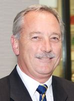
Mr David Adams
Vascular Surgeon
-
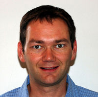
Dr Stuart Barnard
Interventional Radiologist
-
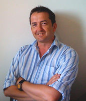
Mr Stuart Caldwell
Vascular Surgeon
-

Dr Daniel Cookson
Interventional Radiologist
-
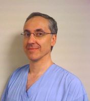
Dr Stewart Hawkins
Interventional Radiologist
-

Dr Giri Mahadevan
Vascular Surgeon
-

Dr Brendon O'Donoghue
Interventional Radiologist
-

Mr Sergei Tchijov
Vascular Surgeon
-
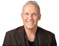
Dr Philip Weeks
Interventional Radiologist
Referral Expectations
If you have an urgent problem requiring immediate surgical assessment you are referred acutely to the Vascular Team where you will initially seen by the junior medical staff in Emergency Care (EC) at Middlemore. They will decide whether you need to be admitted to hospital. Investigations will be performed as required, and the more senior members of the team involved where necessary.
If the problem is not urgent, the GP will write a letter to the Vascular Department requesting an appointment in the outpatient clinic. Waiting times for the clinics depend on the urgency of each case. Urgency is assessed from the information in the GP's referral letter.
When you come to the outpatient clinic you will be seen by a member of the Vascular team who will ask questions about your problem and examine you to try to determine or confirm the diagnosis. This process may also require a number of tests (e.g. blood tests, x-rays, scans etc). Rarely is this done during one clinic visit due to time and will usually require follow-up appointments. Occasionally some tests are arranged even before you are seen at the hospital, to try to speed up the process (vascular ultrasound tests).
Diagnostic tests may include ultrasound, CT, MR and catheter angiography. Interventional Radiologists usually supervise and report these tests and perform all catheter based angiograms. Treatments including angioplasty and stents are sometimes performed by interventional radiologist at the time of the catheter angiogram depending on your clinical status.
Once a diagnosis has been made, the medical staff will discuss treatment with you. In some instances this will mean surgery or catheter angiography. If surgery is advised you will be put on the elective surgical waiting list. Again these waiting lists are ordered according to the urgency and severity of the condition. The steps involved in the surgical process and the likely outcome are usually discussed with you at this time.
In order to minimise the amount of time of that you have to spend in hospital, many surgical departments run a preadmission process. This is usually done through a clinic where you are seen prior to hospital admission. The aim of this clinic is to confirm that you still need to have the planned surgery and that you are currently fit and well enough to undergo the operation. This process usually involves the junior medical staff working in consultation with the anaesthetists, pharmacists, physiotherapists etc. Often the consultant surgeon will also take this opportunity to review your condition.
Outpatient clinics are run at Manukau SuperClinic™.
Fees and Charges Description
There are no charges for services to public patients if you are lawfully in New Zealand and meet one of the Eligibility Directions specified criteria set by the Ministry of Health. If you do not meet the criteria, you will be required to pay for the full costs of any medical treatment you receive during your stay.
To check whether you meet the specified eligibility criteria, visit the Ministry of Health website.
For any applicable charges, please phone the Accounts Receivable Office on (09) 276 0060.
Procedures / Treatments
Vascular Surgery is a surgical subspecialty that deals with all diseases that can affect the arteries and veins. The exceptions are the coronary arteries (heart) that the Cardiothoracic Surgeons operate on and the cerebral brain arteries (brain arteries) that the Neurosurgeons are responsible for. There are five main areas in which we work: Aortic Surgery Carotid Surgery Peripheral Vascular Disease Renal Access Surgery Venous Surgery Other areas that we are involved in include: Hyperhidrosis Visceral Artery Thoracic Outlet Syndrome Diabetic Foot Vascular Trauma Click on the specific surgery/ disease link above to view more information. Alternatively, under 'Document Downloads' below, click the Vascular Surgery link to view and/or print a document with the surgery/disease information listed above. Vascular Surgery (PDF, 21.2 KB)
Vascular Surgery is a surgical subspecialty that deals with all diseases that can affect the arteries and veins. The exceptions are the coronary arteries (heart) that the Cardiothoracic Surgeons operate on and the cerebral brain arteries (brain arteries) that the Neurosurgeons are responsible for. There are five main areas in which we work: Aortic Surgery Carotid Surgery Peripheral Vascular Disease Renal Access Surgery Venous Surgery Other areas that we are involved in include: Hyperhidrosis Visceral Artery Thoracic Outlet Syndrome Diabetic Foot Vascular Trauma Click on the specific surgery/ disease link above to view more information. Alternatively, under 'Document Downloads' below, click the Vascular Surgery link to view and/or print a document with the surgery/disease information listed above. Vascular Surgery (PDF, 21.2 KB)
Vascular Surgery is a surgical subspecialty that deals with all diseases that can affect the arteries and veins. The exceptions are the coronary arteries (heart) that the Cardiothoracic Surgeons operate on and the cerebral brain arteries (brain arteries) that the Neurosurgeons are responsible for. There are five main areas in which we work:
Other areas that we are involved in include:
- Hyperhidrosis
- Visceral Artery
- Thoracic Outlet Syndrome
- Diabetic Foot
- Vascular Trauma
Click on the specific surgery/ disease link above to view more information. Alternatively, under 'Document Downloads' below, click the Vascular Surgery link to view and/or print a document with the surgery/disease information listed above.
- Vascular Surgery (PDF, 21.2 KB)
The aorta, which is the main artery of the body can change over time (aneurysm) to the point where it can rupture. This is usually a fatal event. This process often occurs to the part of the aorta inside the abdomen below where the kidney arteries have branched off and an aneurysm of this part of the aorta is called an Abdominal Aortic Aneurysm (AAA). Patients who have an incidental finding of an aortic aneurysm should be referred to a vascular surgeon, from there depending on size the patient will be kept under surveillance with a scan at regular intervals. The risk of rupture depends on the diameter of the aorta and once the threshold is reached (about 5cm) elective repair is usually offered. There are now 2 main methods of repair of an AAA: Open Repair The first is what is called 'open repair' whereby an incision is made in the abdomen and the aneurysmal aorta is replaced by a prosthetic graft. Endovascular Aortic Repair The second technique is called Endovascular Aortic Repair (EVAR). In this procedure access to the arteries is gained via the main groin artery (common femoral artery) and with the guidance of a radiology machine, a specially packaged graft is passed up over a wire and deployed inside the aorta to act as a new pathway for the blood to flow through. The aneurysm is depressurised and won’t rupture. Long term follow up with radiological imaging is required if this technique is used. At Middlemore Hospital this is a combined procedure performed with the Interventional Radiologists.
The aorta, which is the main artery of the body can change over time (aneurysm) to the point where it can rupture. This is usually a fatal event. This process often occurs to the part of the aorta inside the abdomen below where the kidney arteries have branched off and an aneurysm of this part of the aorta is called an Abdominal Aortic Aneurysm (AAA). Patients who have an incidental finding of an aortic aneurysm should be referred to a vascular surgeon, from there depending on size the patient will be kept under surveillance with a scan at regular intervals. The risk of rupture depends on the diameter of the aorta and once the threshold is reached (about 5cm) elective repair is usually offered. There are now 2 main methods of repair of an AAA: Open Repair The first is what is called 'open repair' whereby an incision is made in the abdomen and the aneurysmal aorta is replaced by a prosthetic graft. Endovascular Aortic Repair The second technique is called Endovascular Aortic Repair (EVAR). In this procedure access to the arteries is gained via the main groin artery (common femoral artery) and with the guidance of a radiology machine, a specially packaged graft is passed up over a wire and deployed inside the aorta to act as a new pathway for the blood to flow through. The aneurysm is depressurised and won’t rupture. Long term follow up with radiological imaging is required if this technique is used. At Middlemore Hospital this is a combined procedure performed with the Interventional Radiologists.
The aorta, which is the main artery of the body can change over time (aneurysm) to the point where it can rupture. This is usually a fatal event. This process often occurs to the part of the aorta inside the abdomen below where the kidney arteries have branched off and an aneurysm of this part of the aorta is called an Abdominal Aortic Aneurysm (AAA).
Patients who have an incidental finding of an aortic aneurysm should be referred to a vascular surgeon, from there depending on size the patient will be kept under surveillance with a scan at regular intervals. The risk of rupture depends on the diameter of the aorta and once the threshold is reached (about 5cm) elective repair is usually offered.
There are now 2 main methods of repair of an AAA:
Open Repair
The first is what is called 'open repair' whereby an incision is made in the abdomen and the aneurysmal aorta is replaced by a prosthetic graft.
Endovascular Aortic Repair
The second technique is called Endovascular Aortic Repair (EVAR). In this procedure access to the arteries is gained via the main groin artery (common femoral artery) and with the guidance of a radiology machine, a specially packaged graft is passed up over a wire and deployed inside the aorta to act as a new pathway for the blood to flow through. The aneurysm is depressurised and won’t rupture. Long term follow up with radiological imaging is required if this technique is used. At Middlemore Hospital this is a combined procedure performed with the Interventional Radiologists.
The formation of plaque at the origin of internal carotid artery, which is the main blood supply to the brain, can lead to small fragments of blood and plaques (emboli) moving into the brain and causing a stroke. In the operation of Carotid Endarterectomy, the plaque is removed and the artery repaired to greatly reduce the risk of a stroke occurring.
The formation of plaque at the origin of internal carotid artery, which is the main blood supply to the brain, can lead to small fragments of blood and plaques (emboli) moving into the brain and causing a stroke. In the operation of Carotid Endarterectomy, the plaque is removed and the artery repaired to greatly reduce the risk of a stroke occurring.
The formation of plaque at the origin of internal carotid artery, which is the main blood supply to the brain, can lead to small fragments of blood and plaques (emboli) moving into the brain and causing a stroke. In the operation of Carotid Endarterectomy, the plaque is removed and the artery repaired to greatly reduce the risk of a stroke occurring.
When atherosclerotic plaque causes narrowing (stenosis) or blockage (occlusion) of the arteries that supply the legs, this can lead to problems with walking (claudication) or the development of ulcers and gangrene. There are various options to improve the blood supply (revascularisation). Often an artery can be opened up (angioplastied) using wires, catheters and balloons using x-ray guidance. At Middlemore Hospital this work is undertaken by the Vascular Radiologists who have special training in these techniques. When surgery is necessary there are a number of techniques available. If a bypass graft around the blockage is required, often a vein from your own leg is used to act as the connection (conduit) between the artery above and below the blockage.
When atherosclerotic plaque causes narrowing (stenosis) or blockage (occlusion) of the arteries that supply the legs, this can lead to problems with walking (claudication) or the development of ulcers and gangrene. There are various options to improve the blood supply (revascularisation). Often an artery can be opened up (angioplastied) using wires, catheters and balloons using x-ray guidance. At Middlemore Hospital this work is undertaken by the Vascular Radiologists who have special training in these techniques. When surgery is necessary there are a number of techniques available. If a bypass graft around the blockage is required, often a vein from your own leg is used to act as the connection (conduit) between the artery above and below the blockage.
When atherosclerotic plaque causes narrowing (stenosis) or blockage (occlusion) of the arteries that supply the legs, this can lead to problems with walking (claudication) or the development of ulcers and gangrene.
There are various options to improve the blood supply (revascularisation). Often an artery can be opened up (angioplastied) using wires, catheters and balloons using x-ray guidance. At Middlemore Hospital this work is undertaken by the Vascular Radiologists who have special training in these techniques. When surgery is necessary there are a number of techniques available. If a bypass graft around the blockage is required, often a vein from your own leg is used to act as the connection (conduit) between the artery above and below the blockage.
Patients who have kidney failure need to dialyse to clear their blood of products that would normally be cleared by functioning kidneys. There are two main ways to do this: Continuous Ambulatory Peritoneal Dialysis Continuous Ambulatory Peritoneal Dialysis (CAPD) which involves exchanging fluid through a special catheter (Tenkhoff catheter) that goes into the peritoneal cavity (abdomen). This must be done everyday. Haemodialysis The other major form of dialysis is haemodialysis via a needle in a fistula vein or graft. Blood is sucked out from the body and spun through a dialysis machine and then returned to the body via another needle into the fistula. The fistula vein or graft is created by taking a vein and joining it to an artery. This means that the strong pulsatile flow of an artery is directed straight into a vein which dilates and can be easily ‘needled’ by the dialysis nurses. The flow in the so called fistula vein is now much greater so that the blood can be drained off to the dialysis machine and returned without clotting (thrombosing). The increased flow in the fistula vein causes vibrations to develop in the blood which can be heard by listening to the vein with a stethoscope (bruit) or palpated by gently placing fingers on the vein (twill). The increased blood supply and constant needling puts a strain on the veins and fistulas are often maintained by performing angioplasties and corrective surgery.
Patients who have kidney failure need to dialyse to clear their blood of products that would normally be cleared by functioning kidneys. There are two main ways to do this: Continuous Ambulatory Peritoneal Dialysis Continuous Ambulatory Peritoneal Dialysis (CAPD) which involves exchanging fluid through a special catheter (Tenkhoff catheter) that goes into the peritoneal cavity (abdomen). This must be done everyday. Haemodialysis The other major form of dialysis is haemodialysis via a needle in a fistula vein or graft. Blood is sucked out from the body and spun through a dialysis machine and then returned to the body via another needle into the fistula. The fistula vein or graft is created by taking a vein and joining it to an artery. This means that the strong pulsatile flow of an artery is directed straight into a vein which dilates and can be easily ‘needled’ by the dialysis nurses. The flow in the so called fistula vein is now much greater so that the blood can be drained off to the dialysis machine and returned without clotting (thrombosing). The increased flow in the fistula vein causes vibrations to develop in the blood which can be heard by listening to the vein with a stethoscope (bruit) or palpated by gently placing fingers on the vein (twill). The increased blood supply and constant needling puts a strain on the veins and fistulas are often maintained by performing angioplasties and corrective surgery.
Patients who have kidney failure need to dialyse to clear their blood of products that would normally be cleared by functioning kidneys. There are two main ways to do this:
Continuous Ambulatory Peritoneal Dialysis
Continuous Ambulatory Peritoneal Dialysis (CAPD) which involves exchanging fluid through a special catheter (Tenkhoff catheter) that goes into the peritoneal cavity (abdomen). This must be done everyday.
Haemodialysis
The other major form of dialysis is haemodialysis via a needle in a fistula vein or graft. Blood is sucked out from the body and spun through a dialysis machine and then returned to the body via another needle into the fistula.
The fistula vein or graft is created by taking a vein and joining it to an artery. This means that the strong pulsatile flow of an artery is directed straight into a vein which dilates and can be easily ‘needled’ by the dialysis nurses. The flow in the so called fistula vein is now much greater so that the blood can be drained off to the dialysis machine and returned without clotting (thrombosing). The increased flow in the fistula vein causes vibrations to develop in the blood which can be heard by listening to the vein with a stethoscope (bruit) or palpated by gently placing fingers on the vein (twill).
The increased blood supply and constant needling puts a strain on the veins and fistulas are often maintained by performing angioplasties and corrective surgery.
Varicose veins (dilated and tortuous veins that can easily be seen in patient’s legs) is a very common condition. Largely they don’t cause major problems and can be managed by General Practitioners with simple measures such as wearing special stockings. However, occasionally they cause significant symptoms or disfigurement (cosmesis) which may threaten a patient’s employment or cause significant loss of quality of life. Also on occasions they can lead to damage to the skin on the legs and this may predispose patients to the development of ulcers. In these situations, Vascular Surgeons are often asked to see and manage these patients. Surgery is offered to those patients who meet certain criteria and when other treatments such as compression stockings have failed. Unfortunately, because this condition is so common it is only those patients with more severe disease who can be offered surgery.
Varicose veins (dilated and tortuous veins that can easily be seen in patient’s legs) is a very common condition. Largely they don’t cause major problems and can be managed by General Practitioners with simple measures such as wearing special stockings. However, occasionally they cause significant symptoms or disfigurement (cosmesis) which may threaten a patient’s employment or cause significant loss of quality of life. Also on occasions they can lead to damage to the skin on the legs and this may predispose patients to the development of ulcers. In these situations, Vascular Surgeons are often asked to see and manage these patients. Surgery is offered to those patients who meet certain criteria and when other treatments such as compression stockings have failed. Unfortunately, because this condition is so common it is only those patients with more severe disease who can be offered surgery.
Varicose veins (dilated and tortuous veins that can easily be seen in patient’s legs) is a very common condition. Largely they don’t cause major problems and can be managed by General Practitioners with simple measures such as wearing special stockings. However, occasionally they cause significant symptoms or disfigurement (cosmesis) which may threaten a patient’s employment or cause significant loss of quality of life. Also on occasions they can lead to damage to the skin on the legs and this may predispose patients to the development of ulcers. In these situations, Vascular Surgeons are often asked to see and manage these patients. Surgery is offered to those patients who meet certain criteria and when other treatments such as compression stockings have failed. Unfortunately, because this condition is so common it is only those patients with more severe disease who can be offered surgery.
The Vascular Laboratory Service is located within the Vascular outpatients Module 9 at Manukau SuperClinic. There are three Vascular Sonographers and they provide a service with surgeons and the Vascular Nurse Specialist. If for any reason you are unable to attend your appointment please inform us so another patient may benefit from your appointment. Your appointment may take anywhere between thirty minutes and one and a half hours, depending on the type of scan you require.
The Vascular Laboratory Service is located within the Vascular outpatients Module 9 at Manukau SuperClinic. There are three Vascular Sonographers and they provide a service with surgeons and the Vascular Nurse Specialist. If for any reason you are unable to attend your appointment please inform us so another patient may benefit from your appointment. Your appointment may take anywhere between thirty minutes and one and a half hours, depending on the type of scan you require.
The Vascular Laboratory Service is located within the Vascular outpatients Module 9 at Manukau SuperClinic. There are three Vascular Sonographers and they provide a service with surgeons and the Vascular Nurse Specialist.
If for any reason you are unable to attend your appointment please inform us so another patient may benefit from your appointment. Your appointment may take anywhere between thirty minutes and one and a half hours, depending on the type of scan you require.
If you are a patient with the Vascular team, you may be seen by a Vascular Nurse Specialist who undertakes case management of all vascular patients and has responsibility for ongoing education of nursing staff and patients. The Vascular Nurse Specialist operates a nurse-run clinic seeing follow-up vascular patients and assessing vascular disease states.
If you are a patient with the Vascular team, you may be seen by a Vascular Nurse Specialist who undertakes case management of all vascular patients and has responsibility for ongoing education of nursing staff and patients. The Vascular Nurse Specialist operates a nurse-run clinic seeing follow-up vascular patients and assessing vascular disease states.
If you are a patient with the Vascular team, you may be seen by a Vascular Nurse Specialist who undertakes case management of all vascular patients and has responsibility for ongoing education of nursing staff and patients. The Vascular Nurse Specialist operates a nurse-run clinic seeing follow-up vascular patients and assessing vascular disease states.
Interventional Radiologists are Specialist Radiologists who specialise in minimally invasive, targeted treatments. Interventional Radiologists offer in-depth knowledge of the minimally invasive treatments coupled with diagnostic and clinical expertise. They use x-rays, MRI and other imaging to advance a catheter in the body, usually in an artery, to treat an abnormality within the artery. Angioplasty (ballooning) and stenting of arteries can be used in the legs to treat peripheral arterial disease. Interventional Radiologists pioneered many techniques which have been adapted to be used in many branches of healthcare. Today many conditions that once required surgery can be treated nonsurgically using those techniques often with less risk and faster recovery time when compared with open surgery. At Middlemore Hospital, all of these endovascular techniques are available to be used to advance the care of patients with vascular disease. The Vascular Surgeons and Interventional Radiologists work in close collaboration to determine the best management plan for each individual patient.
Interventional Radiologists are Specialist Radiologists who specialise in minimally invasive, targeted treatments. Interventional Radiologists offer in-depth knowledge of the minimally invasive treatments coupled with diagnostic and clinical expertise. They use x-rays, MRI and other imaging to advance a catheter in the body, usually in an artery, to treat an abnormality within the artery. Angioplasty (ballooning) and stenting of arteries can be used in the legs to treat peripheral arterial disease. Interventional Radiologists pioneered many techniques which have been adapted to be used in many branches of healthcare. Today many conditions that once required surgery can be treated nonsurgically using those techniques often with less risk and faster recovery time when compared with open surgery. At Middlemore Hospital, all of these endovascular techniques are available to be used to advance the care of patients with vascular disease. The Vascular Surgeons and Interventional Radiologists work in close collaboration to determine the best management plan for each individual patient.
Interventional Radiologists are Specialist Radiologists who specialise in minimally invasive, targeted treatments.
Interventional Radiologists offer in-depth knowledge of the minimally invasive treatments coupled with diagnostic and clinical expertise.
They use x-rays, MRI and other imaging to advance a catheter in the body, usually in an artery, to treat an abnormality within the artery.
Angioplasty (ballooning) and stenting of arteries can be used in the legs to treat peripheral arterial disease. Interventional Radiologists pioneered many techniques which have been adapted to be used in many branches of healthcare.
Today many conditions that once required surgery can be treated nonsurgically using those techniques often with less risk and faster recovery time when compared with open surgery.
At Middlemore Hospital, all of these endovascular techniques are available to be used to advance the care of patients with vascular disease. The Vascular Surgeons and Interventional Radiologists work in close collaboration to determine the best management plan for each individual patient.
The Interventional Radiologists at Counties Manukau DHB are able to offer patients many minimally invasive treatment options. Please click on the links below to find out more information about common interventional radiology procedures: Angiography Arteriovenous Malformations (AVM) Balloon Angioplasty Bleeding Internally Central Venous Access Embolisation Haemodialysis Access Maintenance High Blood Pressure Stent Stent Graft Thrombloysis TIPS (Transjugular Intrahepatic Portosystemic Shunt) Varicose Vein Treatment Vena Cava Filter
The Interventional Radiologists at Counties Manukau DHB are able to offer patients many minimally invasive treatment options. Please click on the links below to find out more information about common interventional radiology procedures: Angiography Arteriovenous Malformations (AVM) Balloon Angioplasty Bleeding Internally Central Venous Access Embolisation Haemodialysis Access Maintenance High Blood Pressure Stent Stent Graft Thrombloysis TIPS (Transjugular Intrahepatic Portosystemic Shunt) Varicose Vein Treatment Vena Cava Filter
The Interventional Radiologists at Counties Manukau DHB are able to offer patients many minimally invasive treatment options.
Please click on the links below to find out more information about common interventional radiology procedures:
Visiting Hours
Ward 8 & Ward 9, Middlemore Hospital
Visiting times are between 8am and 8pm. Visitors are restricted to two at a time.
Children are welcome, but must be supervised by an adult.
If you would like to visit a patient outside of visiting hours (2pm-8pm), please contact the Charge Nurse to discuss. Call (09) 276 0000 and ask to speak to:
Ward 8: Simileshni Kumar, Charge Nurse
Ward 9: Kate Henderson, Charge Nurse
34 E: Helen Bretherton, Charge Nurse
34 N: Manpreet Brar, Charge Nurse
Manukau Surgery Centre
Visiting times are between 11am and 8pm. Visitors are restricted to two at a time.
Children are welcome, but must be supervised by an adult.
If you would like to visit a patient outside of visiting hours (11am-8pm), please contact the Charge Nurse to discuss. Call (09) 277 1660 and ask to speak to:
MSC 2: Shamim Changin, Charge Nurse
MSC 1: Jo Friend, Charge Nurse
Other
For information about parking, refreshments, travel directions, transport, accommodation, support services and maps, click the link below for the required location information:
Website
Contact Details
Manukau Surgery Centre
South Auckland
-
Phone
FREEPHONE 0800 266 513
Website
Ph: (09) 277 1608 (Theatre Unit)
Ph: (09) 277 1655 (Ward Enquiries)
Ph: (09) 250 8034 (Rito Unit)
Email: customerservice@cmdhb.org.nz
Outpatient appointments
& surgical booking enquiries
Ph (09) 277 1660
901 Great South Road
Manurewa
Auckland
Street Address
901 Great South Road
Manurewa
Auckland
Postal Address
General Surgery Department
Private Bag 93311
Ōtāhuhu
Auckland 1640
Middlemore Hospital
South Auckland
-
Phone
(09) 276 0000 or FREEPHONE 0800 266 513
Email
Website
Manukau SuperClinic™
South Auckland
-
Phone
(09) 277 1660 or FREEPHONE 0800 266 513
Email
Website
Was this page helpful?
This page was last updated at 9:56AM on August 8, 2023. This information is reviewed and edited by Vascular Surgery Service | Counties Manukau | Te Whatu Ora.

