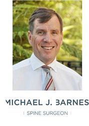
Michael Barnes
Spine Surgeon
PO Box 8482
Symonds Street
Auckland 1150
New Zealand

Michael Barnes - Spine and Orthopaedic Surgeon

Postal Address
Contact Details
Phone (09) 520 0208 or Freephone No: 0800 724 284
Fax (09) 520 0214
Email practicemanager@michaelbarnes.co.nz
Healthlink EDI: mjbarnes
Practice manager: Jennifer Barraclough
POSTERIOR CORRECTION & FUSION FOR SCOLIOSIS
Key Points
- The exact cause of idiopathic scoliosis is unknown but probably has a genetic basis in most patients.
- Surgery is generally recommended when a curve is going to progress beyond 40° to 45°.
- Surgery nearly always involves insertion of implants (screws and rods) onto the spine and fusion (joining one vertebra to the next, bone-to-bone).
- The incision is over the length of the curve and generally heals to a fine line but sometimes spreads slightly at the base of the neck or the bottom of the spine.
- On average, patients spend five nights in hospital after surgery and leave hospital when their pain is under control with tablets, when they are able to get in and out of bed and up and down stairs.
- Patients usually return to school at about four weeks, on half-days for the first two weeks.
- Considering the extent of the surgery, complications are very rare.
- Injury to a spinal nerve or the spinal cord occurs in less than 3 patients out of 1000.
Frequently Asked Questions
Will the rods stop the spine growing?
This spine will not grow significantly over the length of the rods. However, after the age of 10 years 80% of spinal growth is complete and at that the usual time of surgery for adolescent idiopathic scoliosis there is generally only a few millimetres of spinal growth left. This growth simply deforms the spine and shortens it rather than adding to height. The unfused part of the spine and the lower extremities keep growing. Therefore, patients generally end up taller with surgery than without surgery.
Do the rods need to be removed?
It is extremely unusual for the rods or a portion of the rods to require removal. This would only occur if there is an infection or if the rods are prominent under the skin and with current techniques that virtually never occurs.
Will other surgery be required later?
The surgery aims to straighten the spine and protect the unfused parts of the spine above and below the rods. It is very unusual for further surgery to be required. Straightening the major curve at a younger age can at times prevent the need for a longer fusion when the patient is older.
Posterior Correction & Fusion for Scoliosis
General Information
Scoliosis is a lateral (sideways) curvature of the spine. There are multiple causes for scoliosis. The three major categories are:-
A) Neuromuscular (a condition of nerve or muscle such as poliomyelitis)
B) Congenital (the vertebrae are abnormally formed at birth e.g. half a vertebra or
vertebrae fused together)
C) Idiopathic scoliosis (affects otherwise normal children or teenagers, most commonly
females).
Most often patients with neuromuscular scoliosis are in a wheelchair. Congenital scoliosis is apparent on the X-ray because of abnormal vertebrae.
The exact cause of idiopathic scoliosis is unknown but probably has a genetic basis in most patients. Idiopathic scoliosis tends to progress (worsen) during growth. It may progress after the end of growth if moderate (greater than 45°) or severe.
Exercise has not been shown conclusively slow the progression of scoliosis. In selected cases a brace may slow the progression of scoliosis but does not correct it.
Approximately one third of patients with scoliosis have significant pain, although not usually severe. This is usually, although not always, helped by surgery. The primary aim of the surgery is to correct the curve and stop it progressing.
Surgery is generally recommended when a curve is going to progress beyond 40° to 45°.
Surgery nearly always involves insertion of implants (screws and rods) into the spine and fusion (joining one vertebra to the next, bone-to-bone).
The most common surgical procedure involves making an incision over the back of the spine (posterior approach).
Surgical Procedure
Surgery usually takes 4 to 6 hours. The incision is over the length of the curve and generally heals to a fine line but sometimes spreads slightly at the base of the neck or the bottom of the spine.
Surgery usually corrects the curvature by about two thirds and the rib prominence by about two thirds.
Occasionally segments of rib are removed to further correct a severe rib prominence. These grow back later.
Surgery involves inserting screws into the vertebrae (or sometimes attaching hooks) and joining the screws to two rods shaped to the normal contour of the spine to straighten the curve.
Bone is removed from the spine and sometimes the back of the pelvis (hip) and laid in the joints on the back of the spine and over the back of the spine to create a fusion. Sometimes bone graft substitutes are used in addition to bone from the spine to assist fusion.
In large, rigid curves there is a small risk the spinal cord may be stretched and spinal cord monitoring or a wake-up test are performed to ensure this has not occurred.
The wake-up test is performed towards the end of the procedure once the spine has been straightened. The anaesthetic is lightened while the pain killing drugs are working and the patient is asked to move the toes and feet then the anaesthetic is deepened again. This is a safe, routine procedure and patients have no recollection of it later.
Post Operative Treatment
Patients rest in bed the day after the surgery.
For the first two to three days pain relief is from a self-regulated Morphine pump following by oral analgesics (painkillers).
On the second day after surgery the surgical drain is removed and the patient stands out of bed.
On the third day after surgery patients are usually walking to the toilet with assistance and the urinary catheter is removed on the third or fourth day.
On average, patients spend five nights in hospital after surgery and leave hospital when their pain is under control with tablets, when they are able to get in and out of bed and up and down stairs.
There is no particular need to rest after surgery but obviously patients will feel tired for the first several weeks.
Over the six weeks following surgery patients ease back into normal activity. The only exercise necessary is a progressive increase in walking which should be performed regularly. If desired, swimming can commence two weeks following surgery.
Patients usually return to school at about four weeks, on half-days for the first two weeks.
Return to sport usually takes four to six months.
Complications
Considering the extent of the surgery, complications are very rare.
Infection occurs in 1% of patients and is often very low grade and may not show up for many months.
In 1% to 2% of patients the bone does not fuse, leading to pain or breakage of the rods – further surgery may be required.
1% of patients have significant ongoing pain caused by the surgery.
Injury to a spinal nerve or the spinal cord occurs in less than 3 patients out of 1000.
In a very small proportion of patients the spine above or below the rods curves and requires surgery later.
https://www.healthpoint.co.nz/private/orthopaedics/michael-barnes-spine-and-orthopaedic-surgeon/