Central Auckland > Private Hospitals & Specialists > Southern Cross Hospitals >
Southern Cross Gillies Hospital - Otolaryngology, Head & Neck Surgery
Private Surgical Service, ENT/ Head & Neck Surgery
Description
Gillies, a Southern Cross Hospital, is located in the heart of Epsom, Auckland.
The hospital prides itself on being a trusted private surgical hospital with highly skilled professionals providing accessible family-centred care. We value Excellence, Respect, Teamwork and Fairness.
Gillies Hospital has been designed with a focus on short stay surgery, and provides a friendly and highly professional environment.
The hospital's four operating rooms are complemented by 16 inpatient beds and a same day unit.
Consultants
-

Dr Jacqui Allen
Otolaryngologist
-

Dr Stephen Ball
Otolaryngologist
-

Dr Colin Barber
Otolaryngologist
-
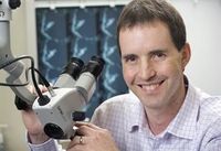
Mr Colin Brown
Otolaryngologist
-
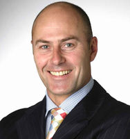
Mr John Chaplin
Otolaryngologist, Head & Neck Surgeon
-

Mr Andrew Cho
Otolaryngologist, Head & Neck Surgeon
-

Dr Melanie Collins
Otolaryngologist
-

Mr Michael Davison
Otolaryngologist
-

Mr Bren Dorman
Otolaryngologist
-

Professor Richard Douglas
Otolaryngologist
-

Mr Joseph Earles
Otolaryngologist
-

Mr David Flint
Otolaryngologist
-
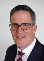
Mr Francis Hall
Otolaryngologist, Head & Neck Surgeon
-

Dr Raymond Kim
Otolaryngologist
-

Mr Nick Lilic
Otolaryngologist
-

Associate Professor Murali Mahadevan
Otolaryngologist, Paediatric Head & Neck Surgeon
-
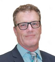
Mr Nick McIvor
Otolaryngologist, Head & Neck Surgeon
-
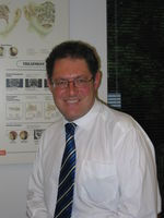
Mr David Mills
Otolaryngologist
-
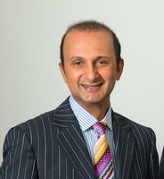
Mr Salil Nair
Otolaryngologist
-

Dr Michel Neeff
Otolaryngologist
-

Mr Sumit Samant
Otolaryngologist
-

Mr Angus Shao
Otolaryngologist, Head & Neck Surgeon
-

Mr Hamish Sillars
Otolaryngologist
-

Dr Ed Toll
Otolaryngologist
-

Dr Graeme van der Meer
Otolaryngologist
-

Dr Veronika van Dijck
Otolaryngologist
-

Mr David Vokes
Otolaryngologist
-

Dr Michelle Wong
Otolaryngologist
Procedures / Treatments
Your adenoids may be removed as part of a tonsillectomy. This operation is also performed through your mouth.
Your adenoids may be removed as part of a tonsillectomy. This operation is also performed through your mouth.
A tiny camera attached to a long tube is inserted through your nose or mouth and passed down through the airways into your lungs. This allows the surgeon to make a diagnosis either by seeing directly what is causing the problem or by taking a small tissue (biopsy) or lung secretion sample.
A tiny camera attached to a long tube is inserted through your nose or mouth and passed down through the airways into your lungs. This allows the surgeon to make a diagnosis either by seeing directly what is causing the problem or by taking a small tissue (biopsy) or lung secretion sample.
An incision (cut) is made behind your ear and the skin pulled back exposing the mastoid bone. A hole is drilled through this bone to expose the cochlear. The electrodes of the cochlear implant are inserted into the cochlear while the receiver part of the implant is embedded into the skull just underneath the skin. The skin is then replaced back over the implant.
An incision (cut) is made behind your ear and the skin pulled back exposing the mastoid bone. A hole is drilled through this bone to expose the cochlear. The electrodes of the cochlear implant are inserted into the cochlear while the receiver part of the implant is embedded into the skull just underneath the skin. The skin is then replaced back over the implant.
A tiny camera attached to a tube (endoscope) is inserted into your nose. Very small instruments can be passed through the endoscope and used to remove small pieces of bone and soft tissue. This opens up the ventilation and drainage pathways in the outer wall of your nose.
A tiny camera attached to a tube (endoscope) is inserted into your nose. Very small instruments can be passed through the endoscope and used to remove small pieces of bone and soft tissue. This opens up the ventilation and drainage pathways in the outer wall of your nose.
This operation is performed through the ear canal. A hole is made in the eardrum and the middle ear drained. A small hollow tube (grommet) is placed in the eardrum hole which allows air into the middle ear.
This operation is performed through the ear canal. A hole is made in the eardrum and the middle ear drained. A small hollow tube (grommet) is placed in the eardrum hole which allows air into the middle ear.
A tiny camera attached to a long tube is inserted into your mouth and passed down through your pharynx into your oesophagus. This allows the surgeon to make a diagnosis either by seeing directly what is causing the problem or by taking a small tissue sample (biopsy).
A tiny camera attached to a long tube is inserted into your mouth and passed down through your pharynx into your oesophagus. This allows the surgeon to make a diagnosis either by seeing directly what is causing the problem or by taking a small tissue sample (biopsy).
Nasal polyps are removed by inserting small instruments through your nostrils which can grasp and cut out the polyps.
Nasal polyps are removed by inserting small instruments through your nostrils which can grasp and cut out the polyps.
Small cuts (incisions) are made either on the inside or outside (in the creases) of the nose. Excess bone and/or cartilage are removed and the nose reshaped.
Small cuts (incisions) are made either on the inside or outside (in the creases) of the nose. Excess bone and/or cartilage are removed and the nose reshaped.
Tonsils are removed in an operation performed through your mouth. The tissue between your tonsils and throat is cut and your tonsils removed.
Tonsils are removed in an operation performed through your mouth. The tissue between your tonsils and throat is cut and your tonsils removed.
This operation repositions the nasal septum and is performed entirely within your nose so that there are no external cuts made on your face.
This operation repositions the nasal septum and is performed entirely within your nose so that there are no external cuts made on your face.
A tiny camera attached to a tube (laryngoscope) is inserted into your mouth and down your throat. This allows the surgeon to make a diagnosis either by seeing directly what is causing the problem or by taking a small tissue sample (biopsy).
A tiny camera attached to a tube (laryngoscope) is inserted into your mouth and down your throat. This allows the surgeon to make a diagnosis either by seeing directly what is causing the problem or by taking a small tissue sample (biopsy).
This operation is performed through the ear canal with an operating microscope. An incision (cut) is made in the middle ear, the small bones of the middle ear are identified and the stapes is removed. A prosthesis is inserted to transmit sound and the wound is closed.
This operation is performed through the ear canal with an operating microscope. An incision (cut) is made in the middle ear, the small bones of the middle ear are identified and the stapes is removed. A prosthesis is inserted to transmit sound and the wound is closed.
This operation is performed through the ear canal with an operating microscope. An incision (cut) is made in the middle ear to view the perforated eardrum, the ear drum is elevated away from the ear canal and lifted forward. A graft of tissue closes the perforation, and the ear is stitched together.
This operation is performed through the ear canal with an operating microscope. An incision (cut) is made in the middle ear to view the perforated eardrum, the ear drum is elevated away from the ear canal and lifted forward. A graft of tissue closes the perforation, and the ear is stitched together.
This operation is performed through the mouth to remove the uvula and the pharyngeal arches and partial removal of the soft palate. Performed as a part of obstructive sleep apnea surgery, the tonsils may also be removed.
This operation is performed through the mouth to remove the uvula and the pharyngeal arches and partial removal of the soft palate. Performed as a part of obstructive sleep apnea surgery, the tonsils may also be removed.
This operation is performed through the mouth to remove the uvula and the pharyngeal arches and partial removal of the soft palate. Performed as a part of obstructive sleep apnea surgery, the tonsils may also be removed.
An incision (cut) is made behind the ear and the air containing space (called the mastoid) is opened. A cortical mastoidectomy involves clearing out diseased tissue in the mastoid cells leaving the ear canal alone. A modified radical mastoidectomy involves joining the spaces of the middle ear canal and the mastoid together to provide an ear that is safe and dry, and hopefully retain some functional hearing.
An incision (cut) is made behind the ear and the air containing space (called the mastoid) is opened. A cortical mastoidectomy involves clearing out diseased tissue in the mastoid cells leaving the ear canal alone. A modified radical mastoidectomy involves joining the spaces of the middle ear canal and the mastoid together to provide an ear that is safe and dry, and hopefully retain some functional hearing.
Refreshments
Tea and Coffee making facilities only on site.
Travel Directions
Click here for map guided travel directions
Public Transport
The Auckland Transport website is a good resource to plan your public transport options.
Parking
A car park is located on the left of the hospital as you enter (off Gillies Ave).
Pharmacy
Contact Details
Southern Cross Gillies Hospital
Central Auckland
For consultant contact details see under "Private Services" on the consultant's profile page.
160 Gillies Avenue
Epsom
Auckland 1023
Street Address
160 Gillies Avenue
Epsom
Auckland 1023
Postal Address
P O Box 99018
Newmarket
Auckland 1149
Was this page helpful?
This page was last updated at 9:37AM on April 3, 2024. This information is reviewed and edited by Southern Cross Gillies Hospital - Otolaryngology, Head & Neck Surgery.

