
Hutt, Wellington > Private Hospitals & Specialists >
Boulcott Hospital - General Surgery
Private Surgical Service, General Surgery
Description
Boulcott Hospital is a 29-bed surgical hospital in Lower Hutt offering state-of-the-art facilities, leading surgeons, a comprehensive range of services and quality care.
We perform a wide range of general surgical procedures.
Consultants
-
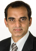
Mr Atul Dhabuwala
General Surgeon
-
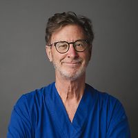
Mr John Groom
General Surgeon
-
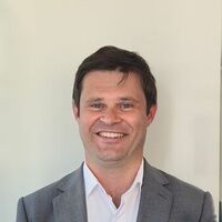
Mr Thomas Morgan
General Surgeon
-
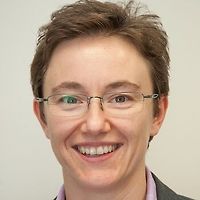
Dr Aleksandra Popadich
General Surgeon
-
Mr Steve Purchas
General Surgeon
Ages
Adult / Pakeke, Child / Tamariki, Older adult / Kaumātua, Youth / Rangatahi
Fees and Charges Categorisation
Fees apply
Fees and Charges Description
Click on the link for information about fees and accounts
Languages Spoken
English
Services Provided
Laparoscopic: several small incisions (cuts) are made in the lower right abdomen (stomach) and a narrow tube with a tiny camera attached (laparoscope) in inserted. This allows the surgeon a view of the appendix and, by inserting small surgical instruments through the other cuts, the appendix can be removed. Open: an incision is made in the lower right abdomen and the appendix removed.
Laparoscopic: several small incisions (cuts) are made in the lower right abdomen (stomach) and a narrow tube with a tiny camera attached (laparoscope) in inserted. This allows the surgeon a view of the appendix and, by inserting small surgical instruments through the other cuts, the appendix can be removed. Open: an incision is made in the lower right abdomen and the appendix removed.
Laparoscopic: several small incisions (cuts) are made in the lower right abdomen (stomach) and a narrow tube with a tiny camera attached (laparoscope) in inserted. This allows the surgeon a view of the appendix and, by inserting small surgical instruments through the other cuts, the appendix can be removed.
Open: an incision is made in the lower right abdomen and the appendix removed.
A small sample of breast tissue is removed and examined under a microscope to see if cancer is present.
A small sample of breast tissue is removed and examined under a microscope to see if cancer is present.
A small sample of breast tissue is removed and examined under a microscope to see if cancer is present.
Simple or Total: all breast tissue, skin and the nipple are surgically removed but the muscles lying under the breast and the lymph nodes are left in place. Modified Radical: all breast tissue, skin and the nipple as well as some lymph tissue are surgically removed. Partial: the breast lump and a portion of other breast tissue (up to one quarter of the breast) as well as lymph tissue are surgically removed. Lumpectomy: the breast lump and surrounding tissue, as well as some lymph tissue, are surgically removed. When combined with radiation treatment, this is known as breast-conserving surgery.
Simple or Total: all breast tissue, skin and the nipple are surgically removed but the muscles lying under the breast and the lymph nodes are left in place. Modified Radical: all breast tissue, skin and the nipple as well as some lymph tissue are surgically removed. Partial: the breast lump and a portion of other breast tissue (up to one quarter of the breast) as well as lymph tissue are surgically removed. Lumpectomy: the breast lump and surrounding tissue, as well as some lymph tissue, are surgically removed. When combined with radiation treatment, this is known as breast-conserving surgery.
Simple or Total: all breast tissue, skin and the nipple are surgically removed but the muscles lying under the breast and the lymph nodes are left in place.
Modified Radical: all breast tissue, skin and the nipple as well as some lymph tissue are surgically removed.
Partial: the breast lump and a portion of other breast tissue (up to one quarter of the breast) as well as lymph tissue are surgically removed.
Lumpectomy: the breast lump and surrounding tissue, as well as some lymph tissue, are surgically removed. When combined with radiation treatment, this is known as breast-conserving surgery.
When a breast has been removed (mastectomy) because of cancer or other disease, it is possible in most cases to reconstruct a breast similar to a natural breast. A breast reconstruction can be performed as part of the breast removal operation or can be performed months or years later. There are two methods of breast reconstruction: one involves using an implant; the other uses tissue taken from another part of your body. There may be medical reasons why one of these methods is more suitable for you or, in other cases, you may be given a choice. Implants A silicone sack filled with either silicone gel or saline (salt water) is inserted underneath the chest muscle and skin. Before being inserted, the skin will sometimes need to be stretched to the required breast size. This is done by placing an empty bag where the implant will finally go, and gradually filling it with saline over weeks or months. The bag is then replaced by the implant in an operation that will probably take 2-3 hours under general anaesthesia (you will sleep through it). You will probably stay in hospital for 2-5 days. Flap reconstruction A flap taken from another part of the body such as your back, stomach or buttocks, is used to reconstruct the breast. This is a more complicated operation than having an implant and may last up to 6 hours and require a 5- to 7-day stay in hospital.
When a breast has been removed (mastectomy) because of cancer or other disease, it is possible in most cases to reconstruct a breast similar to a natural breast. A breast reconstruction can be performed as part of the breast removal operation or can be performed months or years later. There are two methods of breast reconstruction: one involves using an implant; the other uses tissue taken from another part of your body. There may be medical reasons why one of these methods is more suitable for you or, in other cases, you may be given a choice. Implants A silicone sack filled with either silicone gel or saline (salt water) is inserted underneath the chest muscle and skin. Before being inserted, the skin will sometimes need to be stretched to the required breast size. This is done by placing an empty bag where the implant will finally go, and gradually filling it with saline over weeks or months. The bag is then replaced by the implant in an operation that will probably take 2-3 hours under general anaesthesia (you will sleep through it). You will probably stay in hospital for 2-5 days. Flap reconstruction A flap taken from another part of the body such as your back, stomach or buttocks, is used to reconstruct the breast. This is a more complicated operation than having an implant and may last up to 6 hours and require a 5- to 7-day stay in hospital.
When a breast has been removed (mastectomy) because of cancer or other disease, it is possible in most cases to reconstruct a breast similar to a natural breast. A breast reconstruction can be performed as part of the breast removal operation or can be performed months or years later.
There are two methods of breast reconstruction: one involves using an implant; the other uses tissue taken from another part of your body. There may be medical reasons why one of these methods is more suitable for you or, in other cases, you may be given a choice.
Implants
A silicone sack filled with either silicone gel or saline (salt water) is inserted underneath the chest muscle and skin. Before being inserted, the skin will sometimes need to be stretched to the required breast size. This is done by placing an empty bag where the implant will finally go, and gradually filling it with saline over weeks or months. The bag is then replaced by the implant in an operation that will probably take 2-3 hours under general anaesthesia (you will sleep through it). You will probably stay in hospital for 2-5 days.
Flap reconstruction
A flap taken from another part of the body such as your back, stomach or buttocks, is used to reconstruct the breast. This is a more complicated operation than having an implant and may last up to 6 hours and require a 5- to 7-day stay in hospital.
Laparoscopic: several small incisions (cuts) are made in the abdomen and a narrow tube with a tiny camera attached (laparoscope) is inserted. This allows the surgeon a view of the colon and, by inserting small surgical instruments through the other cuts, part or all of the colon can be removed. Open: an abdominal incision is made and part or all of the colon is removed.
Laparoscopic: several small incisions (cuts) are made in the abdomen and a narrow tube with a tiny camera attached (laparoscope) is inserted. This allows the surgeon a view of the colon and, by inserting small surgical instruments through the other cuts, part or all of the colon can be removed. Open: an abdominal incision is made and part or all of the colon is removed.
Laparoscopic: several small incisions (cuts) are made in the abdomen and a narrow tube with a tiny camera attached (laparoscope) is inserted. This allows the surgeon a view of the colon and, by inserting small surgical instruments through the other cuts, part or all of the colon can be removed.
Open: an abdominal incision is made and part or all of the colon is removed.
An opening is made in the skin of the abdomen (stomach) to allow drainage of stools (faeces) from the colon into a collection bag on the outside. This may be temporary to allow time for healing of the colon or, if the entire colon has been removed, it may be permanent.
An opening is made in the skin of the abdomen (stomach) to allow drainage of stools (faeces) from the colon into a collection bag on the outside. This may be temporary to allow time for healing of the colon or, if the entire colon has been removed, it may be permanent.
An opening is made in the skin of the abdomen (stomach) to allow drainage of stools (faeces) from the colon into a collection bag on the outside. This may be temporary to allow time for healing of the colon or, if the entire colon has been removed, it may be permanent.
Gallstones are formed if the gallbladder is not working properly, and the standard treatment is to remove the gallbladder (cholecystectomy). This procedure is usually performed using a laparoscopic (keyhole) approach. Laparoscopic: several small incisions (cuts) are made in the abdomen (stomach) and a narrow tube with a tiny camera attached (laparoscope) is inserted. This allows the surgeon a view of the gallbladder and, by inserting small surgical instruments through the other cuts, the gallbladder can be removed. Open: an abdominal incision is made and the gallbladder removed.
Gallstones are formed if the gallbladder is not working properly, and the standard treatment is to remove the gallbladder (cholecystectomy). This procedure is usually performed using a laparoscopic (keyhole) approach. Laparoscopic: several small incisions (cuts) are made in the abdomen (stomach) and a narrow tube with a tiny camera attached (laparoscope) is inserted. This allows the surgeon a view of the gallbladder and, by inserting small surgical instruments through the other cuts, the gallbladder can be removed. Open: an abdominal incision is made and the gallbladder removed.
Gallstones are formed if the gallbladder is not working properly, and the standard treatment is to remove the gallbladder (cholecystectomy). This procedure is usually performed using a laparoscopic (keyhole) approach.
Laparoscopic: several small incisions (cuts) are made in the abdomen (stomach) and a narrow tube with a tiny camera attached (laparoscope) is inserted. This allows the surgeon a view of the gallbladder and, by inserting small surgical instruments through the other cuts, the gallbladder can be removed.
Open: an abdominal incision is made and the gallbladder removed.
Partial: the diseased part of the stomach is removed and the remaining section is reattached to the oesophagus (food pipe) or small intestine. Total: all of the stomach is removed and the oesophagus is attached directly to the small intestine.
Partial: the diseased part of the stomach is removed and the remaining section is reattached to the oesophagus (food pipe) or small intestine. Total: all of the stomach is removed and the oesophagus is attached directly to the small intestine.
Partial: the diseased part of the stomach is removed and the remaining section is reattached to the oesophagus (food pipe) or small intestine.
Total: all of the stomach is removed and the oesophagus is attached directly to the small intestine.
Haemorrhoids are a condition where the veins under the lining of the anus are congested and enlarged. Less severe haemorrhoids can be managed with simple treatments such as injection or banding which can be performed in the clinic while larger ones will require surgery. Haemorrhoid removal: Haemorrhoidectomy: each haemorrhoid or pile is tied off and then cut away. Stapled Haemorrhoidectomy: a circular stapling device is used to pull the haemorrhoid tissue back into its normal position.
Haemorrhoids are a condition where the veins under the lining of the anus are congested and enlarged. Less severe haemorrhoids can be managed with simple treatments such as injection or banding which can be performed in the clinic while larger ones will require surgery. Haemorrhoid removal: Haemorrhoidectomy: each haemorrhoid or pile is tied off and then cut away. Stapled Haemorrhoidectomy: a circular stapling device is used to pull the haemorrhoid tissue back into its normal position.
Haemorrhoids are a condition where the veins under the lining of the anus are congested and enlarged. Less severe haemorrhoids can be managed with simple treatments such as injection or banding which can be performed in the clinic while larger ones will require surgery.
Haemorrhoid removal:
Haemorrhoidectomy: each haemorrhoid or pile is tied off and then cut away.
Stapled Haemorrhoidectomy: a circular stapling device is used to pull the haemorrhoid tissue back into its normal position.
A hernia exists where part of the abdominal wall is weakened, and the contents of the abdomen push through to the outside. This is most commonly seen in the groin area but can occur in other places. Surgical treatment is usually quite straightforward and involves returning the abdominal contents to the inside and then reinforcing the abdominal wall in some way. Hiatus Hernia: Laparoscopic: several small incisions (cuts) are made in the abdomen (stomach) and a narrow tube with a tiny camera attached (laparoscope) is inserted. Small instruments are inserted through the other cuts, allowing the surgeon to push the hernia (part of the stomach and lower oesophagus that is bulging into the chest) back into position in the abdominal cavity. The hiatus (opening) in the diaphragm (a sheet of muscle between the chest and stomach) is tightened and the stomach is stitched into place. Open: an abdominal incision is made over the hernia and the hernia is pushed back into position in the abdominal cavity. The hiatus (opening in the diaphragm) is tightened and the stomach is stitched into place. Fundoplication: during the above procedures, the top part of the stomach (fundus) may be secured in position by wrapping it around the oesophagus. Inguinal Hernia: Laparoscopic: several small incisions are made in the abdomen and a narrow tube with a tiny camera attached (laparoscope) is inserted. Small instruments are inserted through the other cuts, allowing the surgeon to push the hernia (part of the intestine that is bulging through the abdominal wall) back into its original position. The weakness in the abdominal wall is repaired. Open: an abdominal incision is made and the hernia is pushed back into position. The weakness in the abdominal wall is repaired. Umbilical Hernia: An incision is made underneath the navel (tummy button) and the hernia (part of the intestine that is bulging through the abdominal wall) is pushed back into the abdominal cavity. The weakness in the abdominal wall is repaired. Incisional Hernia: Laparoscopic: several small incisions are made in the abdomen and a narrow tube with a tiny camera attached (laparoscope) is inserted. Small instruments are inserted through the other cuts, allowing the surgeon to push the hernia (part of the intestine that is bulging through the abdominal wall) back into its original position. Open: an abdominal incision is made and the hernia is pushed back into position.
A hernia exists where part of the abdominal wall is weakened, and the contents of the abdomen push through to the outside. This is most commonly seen in the groin area but can occur in other places. Surgical treatment is usually quite straightforward and involves returning the abdominal contents to the inside and then reinforcing the abdominal wall in some way. Hiatus Hernia: Laparoscopic: several small incisions (cuts) are made in the abdomen (stomach) and a narrow tube with a tiny camera attached (laparoscope) is inserted. Small instruments are inserted through the other cuts, allowing the surgeon to push the hernia (part of the stomach and lower oesophagus that is bulging into the chest) back into position in the abdominal cavity. The hiatus (opening) in the diaphragm (a sheet of muscle between the chest and stomach) is tightened and the stomach is stitched into place. Open: an abdominal incision is made over the hernia and the hernia is pushed back into position in the abdominal cavity. The hiatus (opening in the diaphragm) is tightened and the stomach is stitched into place. Fundoplication: during the above procedures, the top part of the stomach (fundus) may be secured in position by wrapping it around the oesophagus. Inguinal Hernia: Laparoscopic: several small incisions are made in the abdomen and a narrow tube with a tiny camera attached (laparoscope) is inserted. Small instruments are inserted through the other cuts, allowing the surgeon to push the hernia (part of the intestine that is bulging through the abdominal wall) back into its original position. The weakness in the abdominal wall is repaired. Open: an abdominal incision is made and the hernia is pushed back into position. The weakness in the abdominal wall is repaired. Umbilical Hernia: An incision is made underneath the navel (tummy button) and the hernia (part of the intestine that is bulging through the abdominal wall) is pushed back into the abdominal cavity. The weakness in the abdominal wall is repaired. Incisional Hernia: Laparoscopic: several small incisions are made in the abdomen and a narrow tube with a tiny camera attached (laparoscope) is inserted. Small instruments are inserted through the other cuts, allowing the surgeon to push the hernia (part of the intestine that is bulging through the abdominal wall) back into its original position. Open: an abdominal incision is made and the hernia is pushed back into position.
A hernia exists where part of the abdominal wall is weakened, and the contents of the abdomen push through to the outside. This is most commonly seen in the groin area but can occur in other places. Surgical treatment is usually quite straightforward and involves returning the abdominal contents to the inside and then reinforcing the abdominal wall in some way.
Hiatus Hernia:
Laparoscopic: several small incisions (cuts) are made in the abdomen (stomach) and a narrow tube with a tiny camera attached (laparoscope) is inserted. Small instruments are inserted through the other cuts, allowing the surgeon to push the hernia (part of the stomach and lower oesophagus that is bulging into the chest) back into position in the abdominal cavity. The hiatus (opening) in the diaphragm (a sheet of muscle between the chest and stomach) is tightened and the stomach is stitched into place.
Open: an abdominal incision is made over the hernia and the hernia is pushed back into position in the abdominal cavity. The hiatus (opening in the diaphragm) is tightened and the stomach is stitched into place.
Fundoplication: during the above procedures, the top part of the stomach (fundus) may be secured in position by wrapping it around the oesophagus.
Inguinal Hernia:
Laparoscopic: several small incisions are made in the abdomen and a narrow tube with a tiny camera attached (laparoscope) is inserted. Small instruments are inserted through the other cuts, allowing the surgeon to push the hernia (part of the intestine that is bulging through the abdominal wall) back into its original position. The weakness in the abdominal wall is repaired.
Open: an abdominal incision is made and the hernia is pushed back into position. The weakness in the abdominal wall is repaired.
Umbilical Hernia:
An incision is made underneath the navel (tummy button) and the hernia (part of the intestine that is bulging through the abdominal wall) is pushed back into the abdominal cavity. The weakness in the abdominal wall is repaired.
Incisional Hernia:
Laparoscopic: several small incisions are made in the abdomen and a narrow tube with a tiny camera attached (laparoscope) is inserted. Small instruments are inserted through the other cuts, allowing the surgeon to push the hernia (part of the intestine that is bulging through the abdominal wall) back into its original position.
Open: an abdominal incision is made and the hernia is pushed back into position.
A range of surgical procedures may be performed on the pancreas, most involving removal of part of the pancreas. Surgery is most commonly required for pancreatic cancer but sometimes for pre-cancerous lesions. Types of surgery include: Whipple procedure: for lesions in the head of the pancreas Distal pancreatectomy: for lesions in the tail or body of the pancreas Total pancreatectomy Surgery may involve: A single large cut in the stomach (open) Using a tiny camera and tools inserted through several small cuts in the stomach (laparoscopic) Robotic arms used by the surgeon to help make the surgery more precise (robot-assisted)
A range of surgical procedures may be performed on the pancreas, most involving removal of part of the pancreas. Surgery is most commonly required for pancreatic cancer but sometimes for pre-cancerous lesions. Types of surgery include: Whipple procedure: for lesions in the head of the pancreas Distal pancreatectomy: for lesions in the tail or body of the pancreas Total pancreatectomy Surgery may involve: A single large cut in the stomach (open) Using a tiny camera and tools inserted through several small cuts in the stomach (laparoscopic) Robotic arms used by the surgeon to help make the surgery more precise (robot-assisted)
A range of surgical procedures may be performed on the pancreas, most involving removal of part of the pancreas. Surgery is most commonly required for pancreatic cancer but sometimes for pre-cancerous lesions.
Types of surgery include:
- Whipple procedure: for lesions in the head of the pancreas
- Distal pancreatectomy: for lesions in the tail or body of the pancreas
- Total pancreatectomy
Surgery may involve:
- A single large cut in the stomach (open)
- Using a tiny camera and tools inserted through several small cuts in the stomach (laparoscopic)
- Robotic arms used by the surgeon to help make the surgery more precise (robot-assisted)
The parathyroid glands are four small glands located in the neck which produce parathyroid hormone, a hormone involved in the regulation of calcium and phosphate levels. Overactivity of one or more of the glands (hyperparathyroidism) results in excessive parathyroid hormone production. Parathyroidectomy is a surgical procedure to remove one or more of the parathyroid glands through an incision (cut) in the front of and at the base of the neck.
The parathyroid glands are four small glands located in the neck which produce parathyroid hormone, a hormone involved in the regulation of calcium and phosphate levels. Overactivity of one or more of the glands (hyperparathyroidism) results in excessive parathyroid hormone production. Parathyroidectomy is a surgical procedure to remove one or more of the parathyroid glands through an incision (cut) in the front of and at the base of the neck.
The parathyroid glands are four small glands located in the neck which produce parathyroid hormone, a hormone involved in the regulation of calcium and phosphate levels. Overactivity of one or more of the glands (hyperparathyroidism) results in excessive parathyroid hormone production.
Parathyroidectomy is a surgical procedure to remove one or more of the parathyroid glands through an incision (cut) in the front of and at the base of the neck.
This is a surgical procedure to remove part or all of the parotid gland, which is the largest of the salivary glands and is located in front of and just below the ear. This surgery is most commonly done to remove tumours, which can be benign (non-cancerous) or malignant (cancerous). It may also be performed for chronic infections or other gland problems. Special care is taken during the surgery to protect the facial nerve, which runs through the parotid gland and controls movement of the face.
This is a surgical procedure to remove part or all of the parotid gland, which is the largest of the salivary glands and is located in front of and just below the ear. This surgery is most commonly done to remove tumours, which can be benign (non-cancerous) or malignant (cancerous). It may also be performed for chronic infections or other gland problems. Special care is taken during the surgery to protect the facial nerve, which runs through the parotid gland and controls movement of the face.
This is a surgical procedure to remove part or all of the parotid gland, which is the largest of the salivary glands and is located in front of and just below the ear.
This surgery is most commonly done to remove tumours, which can be benign (non-cancerous) or malignant (cancerous). It may also be performed for chronic infections or other gland problems.
Special care is taken during the surgery to protect the facial nerve, which runs through the parotid gland and controls movement of the face.
Laparoscopic: several small incisions (cuts) are made in the abdomen (stomach) and a narrow tube with a tiny camera attached (laparoscope) is inserted. This allows the surgeon to view the rectum and, by inserting small surgical instruments through the other cuts, part or all of the rectum can be removed. Open: an abdominal incision is made and part or all of the rectum removed.
Laparoscopic: several small incisions (cuts) are made in the abdomen (stomach) and a narrow tube with a tiny camera attached (laparoscope) is inserted. This allows the surgeon to view the rectum and, by inserting small surgical instruments through the other cuts, part or all of the rectum can be removed. Open: an abdominal incision is made and part or all of the rectum removed.
Laparoscopic: several small incisions (cuts) are made in the abdomen (stomach) and a narrow tube with a tiny camera attached (laparoscope) is inserted. This allows the surgeon to view the rectum and, by inserting small surgical instruments through the other cuts, part or all of the rectum can be removed.
Open: an abdominal incision is made and part or all of the rectum removed.
There are three large pairs of glands (parotid, sublingual and submandibular) in your mouth that produce saliva which helps break down food as part of the digestion process. Salivary gland surgery involves the removal of one or more of the salivary glands for reasons including: tumours (benign or cancerous), chronic infections or blockages, salivary stones or injuries or cysts. Care is taken to avoid damaging nearby nerves, especially those that control facial movement.
There are three large pairs of glands (parotid, sublingual and submandibular) in your mouth that produce saliva which helps break down food as part of the digestion process. Salivary gland surgery involves the removal of one or more of the salivary glands for reasons including: tumours (benign or cancerous), chronic infections or blockages, salivary stones or injuries or cysts. Care is taken to avoid damaging nearby nerves, especially those that control facial movement.
There are three large pairs of glands (parotid, sublingual and submandibular) in your mouth that produce saliva which helps break down food as part of the digestion process.
Salivary gland surgery involves the removal of one or more of the salivary glands for reasons including: tumours (benign or cancerous), chronic infections or blockages, salivary stones or injuries or cysts.
Care is taken to avoid damaging nearby nerves, especially those that control facial movement.
Shave Biopsy: the top layers of skin in the area being investigated are shaved off with a scalpel (surgical knife) for investigation under a microscope. Punch Biopsy: a small cylindrical core of tissue is taken from the area being investigated for examination under a microscope. Excision Biopsy: all of the lesion or area being investigated is cut out with a scalpel for examination under a microscope. Incision Biopsy: part of the lesion is cut out with a scalpel for examination under a microscope.
Shave Biopsy: the top layers of skin in the area being investigated are shaved off with a scalpel (surgical knife) for investigation under a microscope. Punch Biopsy: a small cylindrical core of tissue is taken from the area being investigated for examination under a microscope. Excision Biopsy: all of the lesion or area being investigated is cut out with a scalpel for examination under a microscope. Incision Biopsy: part of the lesion is cut out with a scalpel for examination under a microscope.
Shave Biopsy: the top layers of skin in the area being investigated are shaved off with a scalpel (surgical knife) for investigation under a microscope.
Punch Biopsy: a small cylindrical core of tissue is taken from the area being investigated for examination under a microscope.
Excision Biopsy: all of the lesion or area being investigated is cut out with a scalpel for examination under a microscope.
Incision Biopsy: part of the lesion is cut out with a scalpel for examination under a microscope.
Skin lesions can be divided into two groups: Benign (non-cancerous): e.g. moles, cysts, warts, tags. These may be removed to prevent spreading (warts), stop discomfort if the lesion is being irritated by clothing/jewellery or to improve appearance. Malignant (cancerous): basal cell and squamous cell carcinomas are generally slow growing and unlikely to spread to other parts of the body. Melanoma is a serious skin cancer that can spread to other parts of the body. Urgent removal is recommended. Surgery to remove skin lesions usually involves an office or outpatient visit, local anaesthesia (the area around the scar is numbed by injecting a local anaesthetic) and stitches. You may or may not have a dressing put on the wound and it is important to keep the area dry for 24 hours. Stitches may be removed in 1-2 weeks. You may need to take a few days off work after the surgery.
Skin lesions can be divided into two groups: Benign (non-cancerous): e.g. moles, cysts, warts, tags. These may be removed to prevent spreading (warts), stop discomfort if the lesion is being irritated by clothing/jewellery or to improve appearance. Malignant (cancerous): basal cell and squamous cell carcinomas are generally slow growing and unlikely to spread to other parts of the body. Melanoma is a serious skin cancer that can spread to other parts of the body. Urgent removal is recommended. Surgery to remove skin lesions usually involves an office or outpatient visit, local anaesthesia (the area around the scar is numbed by injecting a local anaesthetic) and stitches. You may or may not have a dressing put on the wound and it is important to keep the area dry for 24 hours. Stitches may be removed in 1-2 weeks. You may need to take a few days off work after the surgery.
Skin lesions can be divided into two groups:
- Benign (non-cancerous): e.g. moles, cysts, warts, tags. These may be removed to prevent spreading (warts), stop discomfort if the lesion is being irritated by clothing/jewellery or to improve appearance.
- Malignant (cancerous): basal cell and squamous cell carcinomas are generally slow growing and unlikely to spread to other parts of the body. Melanoma is a serious skin cancer that can spread to other parts of the body. Urgent removal is recommended.
Surgery to remove skin lesions usually involves an office or outpatient visit, local anaesthesia (the area around the scar is numbed by injecting a local anaesthetic) and stitches. You may or may not have a dressing put on the wound and it is important to keep the area dry for 24 hours. Stitches may be removed in 1-2 weeks. You may need to take a few days off work after the surgery.
The spleen is a soft fleshy organ in the upper left abdomen that is involved in the formation and cleansing of blood. It may need to be removed if it becomes enlarged, has a tumour or cyst, or in the presence of certain blood disorders. Laparoscopic: involves cutting the spleen free from its attachments and removing it through several small incisions (cuts) in the upper left abdomen. Open: an incision is made in the upper left abdomen, the diseased or damaged spleen is then separated from its attachments and removed.
The spleen is a soft fleshy organ in the upper left abdomen that is involved in the formation and cleansing of blood. It may need to be removed if it becomes enlarged, has a tumour or cyst, or in the presence of certain blood disorders. Laparoscopic: involves cutting the spleen free from its attachments and removing it through several small incisions (cuts) in the upper left abdomen. Open: an incision is made in the upper left abdomen, the diseased or damaged spleen is then separated from its attachments and removed.
The spleen is a soft fleshy organ in the upper left abdomen that is involved in the formation and cleansing of blood. It may need to be removed if it becomes enlarged, has a tumour or cyst, or in the presence of certain blood disorders.
Laparoscopic: involves cutting the spleen free from its attachments and removing it through several small incisions (cuts) in the upper left abdomen.
Open: an incision is made in the upper left abdomen, the diseased or damaged spleen is then separated from its attachments and removed.
The thyroid is a gland that sits in the front, and towards the bottom of, your neck. It is responsible for producing a hormone called thyroxin that affects many organs including the heart, muscles and bones. Thyroidectomy is a surgical procedure to remove all or part of the thyroid gland for reasons such as thyroid cancer, goitre (enlarged thyroid), thyroid nodules or overactive thyroid (hyperthyroidism) that doesn't respond to other treatments. A thyroidectomy may be total (removal of the entire thyroid gland) or partial or lobectomy (removal of part of the gland).
The thyroid is a gland that sits in the front, and towards the bottom of, your neck. It is responsible for producing a hormone called thyroxin that affects many organs including the heart, muscles and bones. Thyroidectomy is a surgical procedure to remove all or part of the thyroid gland for reasons such as thyroid cancer, goitre (enlarged thyroid), thyroid nodules or overactive thyroid (hyperthyroidism) that doesn't respond to other treatments. A thyroidectomy may be total (removal of the entire thyroid gland) or partial or lobectomy (removal of part of the gland).
The thyroid is a gland that sits in the front, and towards the bottom of, your neck. It is responsible for producing a hormone called thyroxin that affects many organs including the heart, muscles and bones.
Thyroidectomy is a surgical procedure to remove all or part of the thyroid gland for reasons such as thyroid cancer, goitre (enlarged thyroid), thyroid nodules or overactive thyroid (hyperthyroidism) that doesn't respond to other treatments.
A thyroidectomy may be total (removal of the entire thyroid gland) or partial or lobectomy (removal of part of the gland).
Visiting Hours
Visiting hours are between 8.00am and 8.00pm daily.
Refreshments
For those staying overnight we offer a choice of meals freshly prepared by our resident chef.
Parking
Parking is available for visitors in the grounds of the hospital. Please note this is limited to 90 minutes. If you are staying longer please speak to one of our reception team and they will extend this time limit for you.
Pharmacy
Find the nearest pharmacy here.
Website

Contact Details
Boulcott Hospital
Hutt
-
Phone
(04) 569 7555
Healthlink EDI
boulcott
Email
Website
666 High Street
Boulcott
Lower Hutt
Wellington 5010
Street Address
666 High Street
Boulcott
Lower Hutt
Wellington 5010
Postal Address
PO Box 31 459
Lower Hutt 5040
Wellington
New Zealand
Was this page helpful?
This page was last updated at 8:17AM on September 26, 2025. This information is reviewed and edited by Boulcott Hospital - General Surgery.