
Central Auckland, East Auckland, North Auckland, South Auckland, West Auckland > Private Hospitals & Specialists >
Milford Eye Clinic
Private Service, Ophthalmology
Description
Ophthalmologists are doctors who are trained in the study of eyes. Most will be trained in eye surgery, and may have particular areas of interest or expertise.
Optometrists are not doctors but are trained in testing your vision to assess your need for glasses or contact lenses. Some also test for glaucoma.
For all appointments please contact us on (09) 489 6871.
Consultants
Note: Please note below that some people are not available at all locations.
-
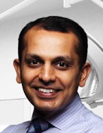
Dr Nadeem Ahmad
Ophthalmologist
Available at Shakespeare Specialist Centre, 181 Shakespeare Road, Milford, Auckland, 9-13 Tamariki Avenue, Ōrewa
-

Dr Rasha Altaie
Ophthalmologist
Available at all locations.
-

Dr Daniel Gosling
Ophthalmologist
Available at all locations.
-

Dr Jo Koppens
Ophthalmologist
Available at all locations.
-

Dr Tahira Malik
Ophthalmologist
Available at all locations.
-
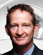
Dr Brian Sloan
Ophthalmologist
Available at all locations.
Fees and Charges Description
Milford Eye Clinic is a Southern Cross Affiliated Provider for Ophthalmology services. These include:
- Cataract surgery
- Chalazion or tarsal cyst removal
- Consultations and assessments
- Corneal cross-linking
- Corneal topography
- Fluorescein angiography
- Fundus photography
- Intravitreal injections
- Laser iridotomy
- Laser trabeculoplasty
- Minor eyelid surgery
- Optical coherence tomography (OCT)
- Photocoagulation of the retina or pan retinal laser
- Pterygium or pinguecula surgery
- Syringing of tear ducts or lacrimal probe
- Visual field tests
- YAG laser capsulotomy
Hours
Shakespeare Specialist Centre, 181 Shakespeare Road, Milford, Auckland
| Mon – Fri | 8:30 AM – 4:30 PM |
|---|
Warkworth Medical Centre, 11 Alnwick Street, Warkworth
As this is a satellite clinic Milford Eye Clinic is open here by appointment only. Please contact us to find out more.
9-13 Tamariki Avenue, Ōrewa
As this is a satellite clinic Milford Eye Clinic is open here by appointment only. Please contact us to find out more.
Procedures / Treatments
These conditions cause distance blur. In myopia, the eye has a resting focus at a near distance so that people will be able to see objects clearly at some point close to them, whilst the distance is blurry. Hyperopia also causes distance blur but often does not become noticeable until the eye loses its ability to change focus, frequently in middle age. The loss of focus for near distance (presbyopia or “aged sight”) is also related to a decreased ability to change focus but only affects reading. Astigmatism causes an image to be blurry at all distances, but does not affect clarity of images unless it is severe. An optometrist or ophthalmologist can test for these conditions. Treatment is usually glasses or contact lenses which are only obtainable through an optometrist or dispensing optician. Laser surgery and other corrective surgical techniques can also be used to change the focus of the eye to give clarity of sight in suitable patients.
These conditions cause distance blur. In myopia, the eye has a resting focus at a near distance so that people will be able to see objects clearly at some point close to them, whilst the distance is blurry. Hyperopia also causes distance blur but often does not become noticeable until the eye loses its ability to change focus, frequently in middle age. The loss of focus for near distance (presbyopia or “aged sight”) is also related to a decreased ability to change focus but only affects reading. Astigmatism causes an image to be blurry at all distances, but does not affect clarity of images unless it is severe. An optometrist or ophthalmologist can test for these conditions. Treatment is usually glasses or contact lenses which are only obtainable through an optometrist or dispensing optician. Laser surgery and other corrective surgical techniques can also be used to change the focus of the eye to give clarity of sight in suitable patients.
These conditions cause distance blur. In myopia, the eye has a resting focus at a near distance so that people will be able to see objects clearly at some point close to them, whilst the distance is blurry. Hyperopia also causes distance blur but often does not become noticeable until the eye loses its ability to change focus, frequently in middle age. The loss of focus for near distance (presbyopia or “aged sight”) is also related to a decreased ability to change focus but only affects reading. Astigmatism causes an image to be blurry at all distances, but does not affect clarity of images unless it is severe. An optometrist or ophthalmologist can test for these conditions. Treatment is usually glasses or contact lenses which are only obtainable through an optometrist or dispensing optician. Laser surgery and other corrective surgical techniques can also be used to change the focus of the eye to give clarity of sight in suitable patients.
Cataracts are the most common age-related problem in eyes. The lens becomes thicker and stiffer and appears yellow and cloudy. Eventually it may turn white, changing the colour of the pupil. A cataract may cause your vision to become fuzzy in a progressive fashion and may also be the cause of disabling glare. Once a cataract affects vision too much, a cataract removal operation is generally advised. This decision is usually made in consultation with an eye specialist. The operation is almost always done under local anaesthetic. Once the cataract has been removed an artificial lens is put in to replace it. It is relatively short in duration and an overnight stay in hospital is not required. Post-operative care consists of eye drops and a check at 1-2 days then after 2-4 weeks.
Cataracts are the most common age-related problem in eyes. The lens becomes thicker and stiffer and appears yellow and cloudy. Eventually it may turn white, changing the colour of the pupil. A cataract may cause your vision to become fuzzy in a progressive fashion and may also be the cause of disabling glare. Once a cataract affects vision too much, a cataract removal operation is generally advised. This decision is usually made in consultation with an eye specialist. The operation is almost always done under local anaesthetic. Once the cataract has been removed an artificial lens is put in to replace it. It is relatively short in duration and an overnight stay in hospital is not required. Post-operative care consists of eye drops and a check at 1-2 days then after 2-4 weeks.
A weakness in one or more of the muscles of the eye will cause the eye to turn or move away from the normal focusing position. This is commonly known as a squint. A squint can be corrected by surgery, or by using glasses. Surgical correction of squint usually involves a general anaesthetic. In the procedure, the muscles involved are repositioned to correct the alignment. It is important to recognise and treat a squint as, if left uncorrected, it can result in permanent impairment of vision.
A weakness in one or more of the muscles of the eye will cause the eye to turn or move away from the normal focusing position. This is commonly known as a squint. A squint can be corrected by surgery, or by using glasses. Surgical correction of squint usually involves a general anaesthetic. In the procedure, the muscles involved are repositioned to correct the alignment. It is important to recognise and treat a squint as, if left uncorrected, it can result in permanent impairment of vision.
Glaucoma is a group of diseases that can damage the eye’s optic nerve and may result in vision loss and blindness. Multiple factors are often important in causing glaucoma, but it is most commonly related to in an increase in pressure in the eye. Symptoms are generally absent until the condition has progressed to an advanced stage. Very occasionally, a rarer form of glaucoma can develop suddenly and symptoms may then include: headaches and aches around the affected eye, seeing halos around lights, sensitivity to light, blurred vision, nausea and vomiting. You may be more likely to develop glaucoma if you: have someone else in your family with glaucoma already have high pressure in your eye have experienced injury to your eye have or have had certain other eye problems have migraine or circulation problems. Glaucoma is more common in people over 50 years of age and more common in women than men. Diagnosis usually comes after consultation with an eye doctor. Signs of glaucoma may also be picked up at an optometrist’s eye examination. The following tests are used to diagnose and monitor glaucoma: Tonometry – measures eye pressure. It is often the first screening test for glaucoma. The eyes are numbed with eye drops and then examined. Dilated eye exam - this is done with an ophthalmoscope (which is a medical instrument that allows the doctor to look through the pupil to the back of the eye).The retina and optic nerve are then examined for any sign of damage. Visual acuity test – test to check distance vision using an eye chart. Visual field test – test to measure side (peripheral) vision. Pachymetry – test to measure the thickness of the cornea. Many other new techniques are emerging to help identify the likelihood of glaucoma and help determine its rate of worsening. Although glaucoma cannot be cured, early treatment can prevent further worsening of the condition and vision loss. Regular eye examinations will need to be continued life-long. Eye drops to decrease eye pressure are the most common early treatment. Surgery may be required, especially if medications are not taking adequate effect. Laser trabeculoplasty, in which a surgeon uses a laser to help the fluid drain from the eye, may be considered in some cases, but has limited effectiveness. More commonly, a trabeculectomy may be performed when other methods have failed to adequately control pressure. This is a medium length operation that makes a new opening for fluid to drain from the eye.
Glaucoma is a group of diseases that can damage the eye’s optic nerve and may result in vision loss and blindness. Multiple factors are often important in causing glaucoma, but it is most commonly related to in an increase in pressure in the eye. Symptoms are generally absent until the condition has progressed to an advanced stage. Very occasionally, a rarer form of glaucoma can develop suddenly and symptoms may then include: headaches and aches around the affected eye, seeing halos around lights, sensitivity to light, blurred vision, nausea and vomiting. You may be more likely to develop glaucoma if you: have someone else in your family with glaucoma already have high pressure in your eye have experienced injury to your eye have or have had certain other eye problems have migraine or circulation problems. Glaucoma is more common in people over 50 years of age and more common in women than men. Diagnosis usually comes after consultation with an eye doctor. Signs of glaucoma may also be picked up at an optometrist’s eye examination. The following tests are used to diagnose and monitor glaucoma: Tonometry – measures eye pressure. It is often the first screening test for glaucoma. The eyes are numbed with eye drops and then examined. Dilated eye exam - this is done with an ophthalmoscope (which is a medical instrument that allows the doctor to look through the pupil to the back of the eye).The retina and optic nerve are then examined for any sign of damage. Visual acuity test – test to check distance vision using an eye chart. Visual field test – test to measure side (peripheral) vision. Pachymetry – test to measure the thickness of the cornea. Many other new techniques are emerging to help identify the likelihood of glaucoma and help determine its rate of worsening. Although glaucoma cannot be cured, early treatment can prevent further worsening of the condition and vision loss. Regular eye examinations will need to be continued life-long. Eye drops to decrease eye pressure are the most common early treatment. Surgery may be required, especially if medications are not taking adequate effect. Laser trabeculoplasty, in which a surgeon uses a laser to help the fluid drain from the eye, may be considered in some cases, but has limited effectiveness. More commonly, a trabeculectomy may be performed when other methods have failed to adequately control pressure. This is a medium length operation that makes a new opening for fluid to drain from the eye.
- have someone else in your family with glaucoma
- already have high pressure in your eye
- have experienced injury to your eye
- have or have had certain other eye problems
- have migraine or circulation problems.
- Tonometry – measures eye pressure. It is often the first screening test for glaucoma. The eyes are numbed with eye drops and then examined.
- Dilated eye exam - this is done with an ophthalmoscope (which is a medical instrument that allows the doctor to look through the pupil to the back of the eye).The retina and optic nerve are then examined for any sign of damage.
- Visual acuity test – test to check distance vision using an eye chart.
- Visual field test – test to measure side (peripheral) vision.
- Pachymetry – test to measure the thickness of the cornea.
This is a complication of diabetes and is caused by small blood vessel damage within the retina of the eye. It commonly affects both eyes and may cause permanent loss of vision. Macular oedema is sometimes also present with diabetic retinopathy. Macular oedema is when fluid leaks into the retina and causes swelling and blurred vision. This may occur at any stage of diabetic retinopathy, but is more common as the disease progresses. There are often no symptoms in the early stages but as the condition progresses vision may begin to become impaired. Often visual loss may be sudden and without warning. This is why it is imperative that at-risk diabetics have frequent eye checks. Poorly controlled diabetes and pregnancy in diabetes are risk factors for developing this condition. Often, first-stage diabetic retinopathy requires no active treatment on the eye but requires stabilisation of diabetes and regular eye examinations. With progressive retinopathy, a laser treatment called the PRP laser can be used. This works by shrinking enlarged blood vessels to prevent further bleeding into the retina. Severe bleeding may require a surgical procedure called a vitrectomy, where blood is surgically removed from the eye. Treatment of macular oedema, if present, is by focal laser treatment. Vision is stabilised by reducing the degree of fluid leakage into the retina. Often more than one treatment is required.
This is a complication of diabetes and is caused by small blood vessel damage within the retina of the eye. It commonly affects both eyes and may cause permanent loss of vision. Macular oedema is sometimes also present with diabetic retinopathy. Macular oedema is when fluid leaks into the retina and causes swelling and blurred vision. This may occur at any stage of diabetic retinopathy, but is more common as the disease progresses. There are often no symptoms in the early stages but as the condition progresses vision may begin to become impaired. Often visual loss may be sudden and without warning. This is why it is imperative that at-risk diabetics have frequent eye checks. Poorly controlled diabetes and pregnancy in diabetes are risk factors for developing this condition. Often, first-stage diabetic retinopathy requires no active treatment on the eye but requires stabilisation of diabetes and regular eye examinations. With progressive retinopathy, a laser treatment called the PRP laser can be used. This works by shrinking enlarged blood vessels to prevent further bleeding into the retina. Severe bleeding may require a surgical procedure called a vitrectomy, where blood is surgically removed from the eye. Treatment of macular oedema, if present, is by focal laser treatment. Vision is stabilised by reducing the degree of fluid leakage into the retina. Often more than one treatment is required.
This is when the retina detaches, meaning it is lifted or separated from its normal position within the eye. An acute retinal detachment requires urgent assessment and appropriate treatment. Unless prompt and effective treatment is given, some forms of retinal detachment may lead to irreversible blindness. Signs and symptoms include: a sudden or gradual increase in floaters, deterioration in vision, cobwebs or specks with the visual field, light flashes in the eye or the appearance of curtains over the visual field. You are more likely to have a retinal detachment if you are very short-sighted or have had an injury or previous surgery to the eye. For minor detachments, a laser or freeze treatment (cryopexy) are used. Both therapies re-attach the retina. For major detachment, surgery will be necessary. A band is often put around the back of the eye to prevent further detachment. Surgical treatment is usually a vitrectomy, where the jelly (vitreous) is removed from the eye. This often involves a hospital stay. It can take several months post-surgery to see the final visual result.
This is when the retina detaches, meaning it is lifted or separated from its normal position within the eye. An acute retinal detachment requires urgent assessment and appropriate treatment. Unless prompt and effective treatment is given, some forms of retinal detachment may lead to irreversible blindness. Signs and symptoms include: a sudden or gradual increase in floaters, deterioration in vision, cobwebs or specks with the visual field, light flashes in the eye or the appearance of curtains over the visual field. You are more likely to have a retinal detachment if you are very short-sighted or have had an injury or previous surgery to the eye. For minor detachments, a laser or freeze treatment (cryopexy) are used. Both therapies re-attach the retina. For major detachment, surgery will be necessary. A band is often put around the back of the eye to prevent further detachment. Surgical treatment is usually a vitrectomy, where the jelly (vitreous) is removed from the eye. This often involves a hospital stay. It can take several months post-surgery to see the final visual result.
Oculoplastic surgery is the sub-specialty part of eye surgery concerned with medical and surgical treatment of eyelid and tear duct problems. In New Zealand's climate, this involves a lot of work treating skin cancers, as well as repair of drooping upper and lower eyelids, skin grafts and blepharoplasty (removal of excess skin in upper or lower eyelids, for functional or cosmetic reasons). Treatment of eyelid changes can be quite different from treatment of skin changes elsewhere, because maintaining the structure and mobility of the eyelids is so important in maintaining the health of the eye. Assessment and treatment of the watery eye is also a large component of oculoplastic practice. Causes of watery eyes include things which increase tear production (such as lashes rubbing the cornea) as well as things which interfere with tear drainage (such as eyelid laxity and tear duct narrowing or blockage). Obviously accurate assessment will allow identification of the most appropriate treatment - medical or surgical. Many small eyelid procedures such as tear duct syringing and eyelid biopsies can be done in the normal clinic appointments. Almost all other procedures are done under local anaesthesia (plus or minus sedation) in the procedure room at Milford Eye Clinic, or in the operating theatres in Shore Surgery (in the same building).
Oculoplastic surgery is the sub-specialty part of eye surgery concerned with medical and surgical treatment of eyelid and tear duct problems. In New Zealand's climate, this involves a lot of work treating skin cancers, as well as repair of drooping upper and lower eyelids, skin grafts and blepharoplasty (removal of excess skin in upper or lower eyelids, for functional or cosmetic reasons). Treatment of eyelid changes can be quite different from treatment of skin changes elsewhere, because maintaining the structure and mobility of the eyelids is so important in maintaining the health of the eye. Assessment and treatment of the watery eye is also a large component of oculoplastic practice. Causes of watery eyes include things which increase tear production (such as lashes rubbing the cornea) as well as things which interfere with tear drainage (such as eyelid laxity and tear duct narrowing or blockage). Obviously accurate assessment will allow identification of the most appropriate treatment - medical or surgical. Many small eyelid procedures such as tear duct syringing and eyelid biopsies can be done in the normal clinic appointments. Almost all other procedures are done under local anaesthesia (plus or minus sedation) in the procedure room at Milford Eye Clinic, or in the operating theatres in Shore Surgery (in the same building).
Oculoplastic surgery is the sub-specialty part of eye surgery concerned with medical and surgical treatment of eyelid and tear duct problems. In New Zealand's climate, this involves a lot of work treating skin cancers, as well as repair of drooping upper and lower eyelids, skin grafts and blepharoplasty (removal of excess skin in upper or lower eyelids, for functional or cosmetic reasons). Treatment of eyelid changes can be quite different from treatment of skin changes elsewhere, because maintaining the structure and mobility of the eyelids is so important in maintaining the health of the eye.
Assessment and treatment of the watery eye is also a large component of oculoplastic practice. Causes of watery eyes include things which increase tear production (such as lashes rubbing the cornea) as well as things which interfere with tear drainage (such as eyelid laxity and tear duct narrowing or blockage). Obviously accurate assessment will allow identification of the most appropriate treatment - medical or surgical.
Many small eyelid procedures such as tear duct syringing and eyelid biopsies can be done in the normal clinic appointments. Almost all other procedures are done under local anaesthesia (plus or minus sedation) in the procedure room at Milford Eye Clinic, or in the operating theatres in Shore Surgery (in the same building).
Public Transport
The Auckland Transport Journey Planner will help you to plan your journey.
Parking
Milford - ample parking at front of building.
Ōrewa - on-street parking (Tamariki Ave).
Warkworth - parking underneath Warkworth Medical Centre, or on-street (Percy & Alnwick streets).
Website

Contact Details
Shakespeare Specialist Centre, 181 Shakespeare Road, Milford, Auckland
North Auckland
-
Phone
(09) 489 6871
Healthlink EDI
milfdeye
Email
Website
Warkworth Medical Centre, 11 Alnwick Street, Warkworth
North Auckland
-
Phone
(09) 489 6871
Healthlink EDI
milfdeye
Email
Website
9-13 Tamariki Avenue, Ōrewa
North Auckland
-
Phone
(09) 489 6871
Healthlink EDI
milfdeye
Email
Website
Was this page helpful?
This page was last updated at 10:08AM on July 10, 2024. This information is reviewed and edited by Milford Eye Clinic.