
Central Auckland, East Auckland, North Auckland, South Auckland, West Auckland > Private Hospitals & Specialists >
Mr Peter Misur - North Shore Hip, Knee & Orthopaedic Trauma Surgeon
Private Service, Orthopaedics
Description
Consultants
-
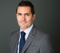
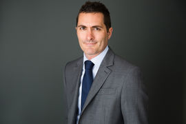
Mr Peter Misur
Hip, Knee & Orthopaedic Trauma Surgeon
Referral Expectations
You need to bring with you to your appointment:
Procedures / Treatments
A hip replacement is a very effective means of alleviating pain from an arthritic or damaged joint. An incision is made on the side of the thigh to allow the surgeon access to the hip joint while causing a minimum of soft tissue disruption. The diseased and damaged parts of the hip joint are removed and replaced with artificial components. Patients can generally start full weight bearing from the day of surgery.
A hip replacement is a very effective means of alleviating pain from an arthritic or damaged joint. An incision is made on the side of the thigh to allow the surgeon access to the hip joint while causing a minimum of soft tissue disruption. The diseased and damaged parts of the hip joint are removed and replaced with artificial components. Patients can generally start full weight bearing from the day of surgery.
A hip replacement is a very effective means of alleviating pain from an arthritic or damaged joint. An incision is made on the side of the thigh to allow the surgeon access to the hip joint while causing a minimum of soft tissue disruption. The diseased and damaged parts of the hip joint are removed and replaced with artificial components. Patients can generally start full weight bearing from the day of surgery.
A hip resurfacing is a form of hip replacement which is generally reserved for younger and more active patients. This procedure differs from a traditional hip replacement in that more of the patient's hip bone is preserved and a metal cap is instead placed to 'resurface' the head of the hip. Patients interested in a resurfacing procedure are welcome to discuss their suitability for this surgery and how the functional outcomes may differ from a traditional hip replacement.
A hip resurfacing is a form of hip replacement which is generally reserved for younger and more active patients. This procedure differs from a traditional hip replacement in that more of the patient's hip bone is preserved and a metal cap is instead placed to 'resurface' the head of the hip. Patients interested in a resurfacing procedure are welcome to discuss their suitability for this surgery and how the functional outcomes may differ from a traditional hip replacement.
A hip resurfacing is a form of hip replacement which is generally reserved for younger and more active patients. This procedure differs from a traditional hip replacement in that more of the patient's hip bone is preserved and a metal cap is instead placed to 'resurface' the head of the hip. Patients interested in a resurfacing procedure are welcome to discuss their suitability for this surgery and how the functional outcomes may differ from a traditional hip replacement.
A knee replacement is used to alleviate the pain of an arthritic joint. Knee replacements may involve part (unicompartmental) or all of the knee joint surfaces depending upon individual patient characteristics. An incision (cut) is made on the front of the knee to allow the surgeon access to the knee joint. The damaged and painful areas of the thigh bone (femur), lower leg bone (tibia) and knee cap (patella) are removed and replaced with artificial components. Rigorous post-operative physiotherapy is prescribed in order to maximise knee function.
A knee replacement is used to alleviate the pain of an arthritic joint. Knee replacements may involve part (unicompartmental) or all of the knee joint surfaces depending upon individual patient characteristics. An incision (cut) is made on the front of the knee to allow the surgeon access to the knee joint. The damaged and painful areas of the thigh bone (femur), lower leg bone (tibia) and knee cap (patella) are removed and replaced with artificial components. Rigorous post-operative physiotherapy is prescribed in order to maximise knee function.
A knee replacement is used to alleviate the pain of an arthritic joint. Knee replacements may involve part (unicompartmental) or all of the knee joint surfaces depending upon individual patient characteristics. An incision (cut) is made on the front of the knee to allow the surgeon access to the knee joint. The damaged and painful areas of the thigh bone (femur), lower leg bone (tibia) and knee cap (patella) are removed and replaced with artificial components. Rigorous post-operative physiotherapy is prescribed in order to maximise knee function.
Long-standing hip and knee replacements may eventually wear. Typically this takes many years to occur and is likely to become less common with the development of more advanced artificial materials. Badly worn components may require revision surgery to replace.
Long-standing hip and knee replacements may eventually wear. Typically this takes many years to occur and is likely to become less common with the development of more advanced artificial materials. Badly worn components may require revision surgery to replace.
Long-standing hip and knee replacements may eventually wear. Typically this takes many years to occur and is likely to become less common with the development of more advanced artificial materials. Badly worn components may require revision surgery to replace.
Knee arthroscopy (key-hole surgery) involves a small incision at the front of the knee for the introduction of a small camera (arthroscope.) This allows the surgeon to look inside the joint, identify problems and, in some cases, make repairs to damaged tissue. This technique is used to address tears of the meniscus joint surface cartilage. Knee ligament injuries can also be reconstructed using these 'key-hole' techniques. The use of key-hole knee arthroscopy, with small skin incisions, means that generally such procedures have a rapid recovery and most patients can return home the same day as their surgery.
Knee arthroscopy (key-hole surgery) involves a small incision at the front of the knee for the introduction of a small camera (arthroscope.) This allows the surgeon to look inside the joint, identify problems and, in some cases, make repairs to damaged tissue. This technique is used to address tears of the meniscus joint surface cartilage. Knee ligament injuries can also be reconstructed using these 'key-hole' techniques. The use of key-hole knee arthroscopy, with small skin incisions, means that generally such procedures have a rapid recovery and most patients can return home the same day as their surgery.
Knee arthroscopy (key-hole surgery) involves a small incision at the front of the knee for the introduction of a small camera (arthroscope.) This allows the surgeon to look inside the joint, identify problems and, in some cases, make repairs to damaged tissue. This technique is used to address tears of the meniscus joint surface cartilage. Knee ligament injuries can also be reconstructed using these 'key-hole' techniques.
The use of key-hole knee arthroscopy, with small skin incisions, means that generally such procedures have a rapid recovery and most patients can return home the same day as their surgery.
The knee has two c-shaped cartilages called 'menisci.' These function as shock-absorbers within the knee, distributing load within the joint. The menisci can be torn as the result of pivoting knee injuries or as part of long-term wear. Sometimes these tears may cause ongoing pain or persisting mechanical catching within the knee. In such cases the tear can be surgically managed with a knee arthroscopy. This is a 'key-hole' procedure in which small incisions are used to introduce a camera and a working instrument into the knee in order to address the tear. The use of key-hole surgery means that patients have a faster recovery and can generally return home the same day as their surgery.
The knee has two c-shaped cartilages called 'menisci.' These function as shock-absorbers within the knee, distributing load within the joint. The menisci can be torn as the result of pivoting knee injuries or as part of long-term wear. Sometimes these tears may cause ongoing pain or persisting mechanical catching within the knee. In such cases the tear can be surgically managed with a knee arthroscopy. This is a 'key-hole' procedure in which small incisions are used to introduce a camera and a working instrument into the knee in order to address the tear. The use of key-hole surgery means that patients have a faster recovery and can generally return home the same day as their surgery.
The knee has two c-shaped cartilages called 'menisci.' These function as shock-absorbers within the knee, distributing load within the joint. The menisci can be torn as the result of pivoting knee injuries or as part of long-term wear. Sometimes these tears may cause ongoing pain or persisting mechanical catching within the knee. In such cases the tear can be surgically managed with a knee arthroscopy. This is a 'key-hole' procedure in which small incisions are used to introduce a camera and a working instrument into the knee in order to address the tear. The use of key-hole surgery means that patients have a faster recovery and can generally return home the same day as their surgery.
The knee joint has four main stabilising ligaments. Not all knee ligament injuries require surgery. Injury to the anterior cruciate ligament (ACL), deep within the knee joint, is often the result of sporting activity. Rupture of the ACL may lead to a sense of knee instability which impairs the return to normal function. An ACL reconstruction can be performed using key-hole (arthroscopic) techniques.
The knee joint has four main stabilising ligaments. Not all knee ligament injuries require surgery. Injury to the anterior cruciate ligament (ACL), deep within the knee joint, is often the result of sporting activity. Rupture of the ACL may lead to a sense of knee instability which impairs the return to normal function. An ACL reconstruction can be performed using key-hole (arthroscopic) techniques.
Service types: Knee surgery, ACL (Anterior cruciate ligament) reconstruction, Soft tissue (muscles, tendons and ligaments).
The knee joint has four main stabilising ligaments. Not all knee ligament injuries require surgery. Injury to the anterior cruciate ligament (ACL), deep within the knee joint, is often the result of sporting activity. Rupture of the ACL may lead to a sense of knee instability which impairs the return to normal function. An ACL reconstruction can be performed using key-hole (arthroscopic) techniques.
A dislocation of the knee cap (patella) may occur on a single occasion and not require any surgical intervention. Some patients may however have repeated knee-cap dislocations in which case surgery will be considered. There are a range of surgical options depending on the anatomy of the individual patient and these include ligament (patello-femoral) reconstruction or a bone re-alignment procedure. Sometimes a combination of procedures may be recommended in order to increase the chance of a successful outcome. Patella stabilisation can be a complex area of orthopaedic surgery, particularly in patients with lax ligaments (sometimes referred to as being 'double-jointed').
A dislocation of the knee cap (patella) may occur on a single occasion and not require any surgical intervention. Some patients may however have repeated knee-cap dislocations in which case surgery will be considered. There are a range of surgical options depending on the anatomy of the individual patient and these include ligament (patello-femoral) reconstruction or a bone re-alignment procedure. Sometimes a combination of procedures may be recommended in order to increase the chance of a successful outcome. Patella stabilisation can be a complex area of orthopaedic surgery, particularly in patients with lax ligaments (sometimes referred to as being 'double-jointed').
Service types: Knee surgery.
A dislocation of the knee cap (patella) may occur on a single occasion and not require any surgical intervention. Some patients may however have repeated knee-cap dislocations in which case surgery will be considered. There are a range of surgical options depending on the anatomy of the individual patient and these include ligament (patello-femoral) reconstruction or a bone re-alignment procedure. Sometimes a combination of procedures may be recommended in order to increase the chance of a successful outcome. Patella stabilisation can be a complex area of orthopaedic surgery, particularly in patients with lax ligaments (sometimes referred to as being 'double-jointed').
Osteotomy is the division of a crooked or bent bone to improve alignment of the limb. These procedures normally involve some form of internal fixation, such as rods or plates, or external fixation which involves external wires and pins to hold the bone. The type of procedure for fixation will be explained when the surgery is planned.
Osteotomy is the division of a crooked or bent bone to improve alignment of the limb. These procedures normally involve some form of internal fixation, such as rods or plates, or external fixation which involves external wires and pins to hold the bone. The type of procedure for fixation will be explained when the surgery is planned.
Osteotomy is the division of a crooked or bent bone to improve alignment of the limb.
These procedures normally involve some form of internal fixation, such as rods or plates, or external fixation which involves external wires and pins to hold the bone. The type of procedure for fixation will be explained when the surgery is planned.
Public Transport
The Auckland Transport Journey Planner will help you to plan your journey.
Parking
Patient parking is provided at 209 Shakepeare Road, Milford and 131 Lincoln Road, Henderson.
Website
Contact Details
209 Shakespeare Road, Milford, Auckland
North Auckland
-
Phone
(09) 486 5456
-
Mobile
021 0287 0854
Healthlink EDI
petermis
Email
Website
131 Lincoln Road, Henderson, Auckland
West Auckland
-
Phone
(09) 486 5456
-
Mobile
021 0287 0854
Healthlink EDI
petermis
Email
Website
Was this page helpful?
This page was last updated at 10:28AM on February 16, 2026. This information is reviewed and edited by Mr Peter Misur - North Shore Hip, Knee & Orthopaedic Trauma Surgeon.