
Canterbury > Private Hospitals & Specialists > Southern Cross Hospitals >
Southern Cross Christchurch Hospital - Paediatric Surgery
Private Surgical Service, Paediatrics
Description
Southern Cross Hospital in Christchurch is the largest hospital within our national network.
Owned by Southern Cross since 1979, the centrally situated hospital campus includes one of the biggest and most advanced private surgical hospitals in the South Island.
The Christchurch hospital campus has seen significant upgrades and new facilities in recent years, and typically provides services to around 9,500 patients each year. Facilities include digital operating theatres, an advanced 'hybrid' operating room, systems for robotically-assisted surgery, advanced digital scanning technologies, consulting facilities and a purpose built endoscopy centre.
Consultants
-
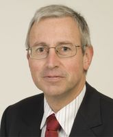
Professor Spencer Beasley
Paediatric Surgeon
-
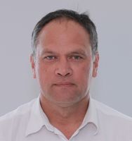
Mr Kiki Maoate
Paediatric Surgeon
Procedures / Treatments
Umbilical Hernia An incision (cut) is made underneath the navel (tummy button) and the hernia (part of the intestine that is bulging through the abdominal wall) is pushed back into the abdominal cavity. The weakness in the abdominal wall is repaired. Inguinal Hernia Laparoscopic: several small incisions are made in the abdomen and a narrow tube with a tiny camera attached (laparoscope) is inserted. Small instruments are inserted through the other cuts, allowing the surgeon to push the hernia (part of the intestine that is bulging through the abdominal wall) back into its original position. The weakness in the abdominal wall is repaired. Open: an abdominal incision is made and the hernia is pushed back into position. The weakness in the abdominal wall is repaired. Herniotomy: an incision is made in a skin fold in the groin and the hernia sac is cut out.
Umbilical Hernia An incision (cut) is made underneath the navel (tummy button) and the hernia (part of the intestine that is bulging through the abdominal wall) is pushed back into the abdominal cavity. The weakness in the abdominal wall is repaired. Inguinal Hernia Laparoscopic: several small incisions are made in the abdomen and a narrow tube with a tiny camera attached (laparoscope) is inserted. Small instruments are inserted through the other cuts, allowing the surgeon to push the hernia (part of the intestine that is bulging through the abdominal wall) back into its original position. The weakness in the abdominal wall is repaired. Open: an abdominal incision is made and the hernia is pushed back into position. The weakness in the abdominal wall is repaired. Herniotomy: an incision is made in a skin fold in the groin and the hernia sac is cut out.
A small incision (cut) is made in the groin on the side of the undescended testicle and the testicle pulled down into the scrotum. Sometimes a small cut will need to be made in the scrotum as well.
A small incision (cut) is made in the groin on the side of the undescended testicle and the testicle pulled down into the scrotum. Sometimes a small cut will need to be made in the scrotum as well.
All lymph nodes (bean-shaped glands that filter harmful agents picked up by the lymphatic system) from the collar bone to the jaw and from the front of the neck to the back are removed, along with the sternocleidomastoid muscle (moves the head from side to side), the spinal accessory nerve (involved in speech, swallowing and some head movements), the submandibular gland (one of the salivary glands) and the internal jugular vein.
All lymph nodes (bean-shaped glands that filter harmful agents picked up by the lymphatic system) from the collar bone to the jaw and from the front of the neck to the back are removed, along with the sternocleidomastoid muscle (moves the head from side to side), the spinal accessory nerve (involved in speech, swallowing and some head movements), the submandibular gland (one of the salivary glands) and the internal jugular vein.
All lymph nodes (bean-shaped glands that filter harmful agents picked up by the lymphatic system) from the collar bone to the jaw and from the front of the neck to the back are removed.
All lymph nodes (bean-shaped glands that filter harmful agents picked up by the lymphatic system) from the collar bone to the jaw and from the front of the neck to the back are removed.
A long, narrow tube with a tiny camera attached (sigmoidoscope) is inserted into the anus and moved through the lower large intestine (bowel). This allows the surgeon a view of the lining of the lower large intestine (sigmoid colon). If necessary, a biopsy (small piece of tissue) may be taken for examination in the laboratory.
A long, narrow tube with a tiny camera attached (sigmoidoscope) is inserted into the anus and moved through the lower large intestine (bowel). This allows the surgeon a view of the lining of the lower large intestine (sigmoid colon). If necessary, a biopsy (small piece of tissue) may be taken for examination in the laboratory.
A fold of tissue (frenum) that attaches to the cheek, lips and/or tongue is surgically removed.
A fold of tissue (frenum) that attaches to the cheek, lips and/or tongue is surgically removed.
Laparoscopic: several small incisions (cuts) are made in the lower right abdomen and a narrow tube with a tiny camera attached (laparoscope) in inserted. This allows the surgeon a view of the appendix and, by inserting small surgical instruments through the other cuts, the appendix can be removed.
Laparoscopic: several small incisions (cuts) are made in the lower right abdomen and a narrow tube with a tiny camera attached (laparoscope) in inserted. This allows the surgeon a view of the appendix and, by inserting small surgical instruments through the other cuts, the appendix can be removed.
The foreskin is pulled away from the body of the penis and cut off, exposing the underlying head of the penis (glans). Stitches may be required to keep the remaining edges of the foreskin in place.
The foreskin is pulled away from the body of the penis and cut off, exposing the underlying head of the penis (glans). Stitches may be required to keep the remaining edges of the foreskin in place.
Shave Biopsy: the top layers of skin in the area being investigated are shaved off with a scalpel (surgical knife) for investigation under a microscope. Punch Biopsy: a small cylindrical core of tissue is taken from the area being investigated for examination under a microscope. Excision Biopsy: all of the lesion or area being investigated is cut out with a scalpel for examination under a microscope. Incision Biopsy: part of the lesion is cut out with a scalpel for examination under a microscope.
Shave Biopsy: the top layers of skin in the area being investigated are shaved off with a scalpel (surgical knife) for investigation under a microscope. Punch Biopsy: a small cylindrical core of tissue is taken from the area being investigated for examination under a microscope. Excision Biopsy: all of the lesion or area being investigated is cut out with a scalpel for examination under a microscope. Incision Biopsy: part of the lesion is cut out with a scalpel for examination under a microscope.
Skin lesions such as cysts and tumours are removed by cutting around and under them with a scalpel.
Skin lesions such as cysts and tumours are removed by cutting around and under them with a scalpel.
A minor surgical procedure is performed to widen the urinary meatus or opening (where the urine exits the body).
A minor surgical procedure is performed to widen the urinary meatus or opening (where the urine exits the body).
A small cut is made in the scrotum and the fluid is drained from the hydrocoele sac (a fluid-filled mass that forms in the scrotum). The sac may either be removed or is folded back behind the testicle.
A small cut is made in the scrotum and the fluid is drained from the hydrocoele sac (a fluid-filled mass that forms in the scrotum). The sac may either be removed or is folded back behind the testicle.
A small cut is made in the scrotum, the cord supplying blood to the testicle is untwisted and both testes are sutured (stitched) to the scrotum to prevent another torsion.
A small cut is made in the scrotum, the cord supplying blood to the testicle is untwisted and both testes are sutured (stitched) to the scrotum to prevent another torsion.
Visiting Hours
Weekdays 8:00 to 20:00
Weekends 8:00 to 20:00
Public Transport
The Christchurch City Council provides good public transport information. See here
Parking
Over 60 parking spaces are provided for patients and visitors.
Pharmacy

Contact Details
Southern Cross Christchurch Hospital
Canterbury
-
Phone
(03) 968 3100
-
Fax
(03) 968 3101
Email
Website
131 Bealey Avenue
Christchurch Central
Christchurch
Canterbury 8013
Street Address
131 Bealey Avenue
Christchurch Central
Christchurch
Canterbury 8013
Postal Address
P.O. Box 21-096,
Edgeware,
Christchurch, 8143
Was this page helpful?
This page was last updated at 2:49PM on April 10, 2024. This information is reviewed and edited by Southern Cross Christchurch Hospital - Paediatric Surgery.