
Waikato > Private Hospitals & Specialists >
River Radiology
Private Service, Radiology, Pregnancy Ultrasound
Today
30 Hood Street, Hamilton
8:00 AM to 5:00 PM.
173 Anglesea Street, Hamilton
8:00 AM to 8:00 PM.
1913 Cambridge Road, Cambridge
8:30 AM to 5:00 PM.
18 Karewa Place, Pūkete, Hamilton
8:30 AM to 5:00 PM.
Description
We are a patient-focused radiology practice offering patients the best possible care in a friendly environment. We strive to solve patients' diagnostic problems and improve patient outcomes by providing imaging excellence and by continuously auditing our practice.
Our services include:
- X-Ray
- Ultrasound - including pregnancy scans
- Cone Beam CT
- MRI
- Guided injections - Corticosteroid, PRP (Platelet-Rich Plasma), Autologous blood, Spinal & Achilles.
Consultants
-
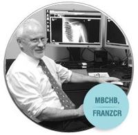
Dr Peter Gendall
Radiologist - Musculoskeletal Imaging
-

Dr Nirushini Sathiyakumar
Radiologist - Musculoskeletal Imaging
-
Dr Pierre Struwig
Neuroradiologist
Referral Expectations
Fees and Charges Description
Click here for our current price list.
Hours
30 Hood Street, Hamilton
8:00 AM to 5:00 PM.
| Mon – Fri | 8:00 AM – 5:00 PM |
|---|
Our services include:
- X-Ray
- Ultrasound - including pregnancy scans
- Cone Beam CT
- MRI
- Guided injections - Corticosteroid, PRP (Platelet-Rich Plasma), Autologous blood, Spinal & Achilles.
173 Anglesea Street, Hamilton
8:00 AM to 8:00 PM.
| Mon – Fri | 8:00 AM – 8:00 PM |
|---|
Our services include:
- X-Ray
- Ultrasound - including pregnancy scans
1913 Cambridge Road, Cambridge
8:30 AM to 5:00 PM.
| Mon – Fri | 8:30 AM – 5:00 PM |
|---|
Our services include:
- X-Ray
- Ultrasound - including pregnancy scans
- Guided injections - Corticosteroid
18 Karewa Place, Pūkete, Hamilton
8:30 AM to 5:00 PM.
| Mon – Fri | 8:30 AM – 5:00 PM |
|---|
Our services include:
- X-Ray
- Ultrasound - including pregnancy scans
- Cone Beam CT
Procedures / Treatments
Here at River Radiology we use a digital x-ray machine that allows us to view your image only seconds after taking it. With state-of-the-art technology we are able to skip the entire processing stage, meaning a speedy process for all our patients. This is the most efficient technology available - you won't see this at every x-ray facility in town!
Here at River Radiology we use a digital x-ray machine that allows us to view your image only seconds after taking it. With state-of-the-art technology we are able to skip the entire processing stage, meaning a speedy process for all our patients. This is the most efficient technology available - you won't see this at every x-ray facility in town!
River Radiology has a Newtom 5G Cone Beam CT, a specialist CT machine. Like all CTs it uses x-rays but an examination on the Cone Beam CT uses only 10% of the radiation delivered by a routine Fan Beam CT! Our Cone Beam differs from the more commonly available dental machines; patients lie down on a moving table to be scanned, this means we can scan elbows, wrists and hands, knees, ankles and feet, and even the cervical spine (neck). The Cone Beam also gets wonderful high resolution scans of face, sinuses, teeth and temporal bones (inner ear).
River Radiology has a Newtom 5G Cone Beam CT, a specialist CT machine. Like all CTs it uses x-rays but an examination on the Cone Beam CT uses only 10% of the radiation delivered by a routine Fan Beam CT! Our Cone Beam differs from the more commonly available dental machines; patients lie down on a moving table to be scanned, this means we can scan elbows, wrists and hands, knees, ankles and feet, and even the cervical spine (neck). The Cone Beam also gets wonderful high resolution scans of face, sinuses, teeth and temporal bones (inner ear).
River Radiology has a Newtom 5G Cone Beam CT, a specialist CT machine. Like all CTs it uses x-rays but an examination on the Cone Beam CT uses only 10% of the radiation delivered by a routine Fan Beam CT!
Our Cone Beam differs from the more commonly available dental machines; patients lie down on a moving table to be scanned, this means we can scan elbows, wrists and hands, knees, ankles and feet, and even the cervical spine (neck). The Cone Beam also gets wonderful high resolution scans of face, sinuses, teeth and temporal bones (inner ear).
River Radiology's state-of-the-art GE 3.0 Tesla scanner performs high-resolution scans in minutes whilst you relax on a comfortable padded table. There is no need for oral sedation because our scan room has been specifically designed to put you at ease. MRI (magnetic resonance imaging) uses a very powerful magnetic field, radio waves and a computer to produce detailed images of almost every internal part of the body. This process is completely safe and involves no harmful radiation whatsoever. Patient Preparation: advise our booking staff if you have any internal metal implants or electronic devices before your MRI scan, eat, drink and take any medication as normal, unless advised otherwise please arrive 15 minutes prior to your scan so you can fill out a safety questionnaire prior to your MRI scan please allow 45-60 minutes for your examination in total.
River Radiology's state-of-the-art GE 3.0 Tesla scanner performs high-resolution scans in minutes whilst you relax on a comfortable padded table. There is no need for oral sedation because our scan room has been specifically designed to put you at ease. MRI (magnetic resonance imaging) uses a very powerful magnetic field, radio waves and a computer to produce detailed images of almost every internal part of the body. This process is completely safe and involves no harmful radiation whatsoever. Patient Preparation: advise our booking staff if you have any internal metal implants or electronic devices before your MRI scan, eat, drink and take any medication as normal, unless advised otherwise please arrive 15 minutes prior to your scan so you can fill out a safety questionnaire prior to your MRI scan please allow 45-60 minutes for your examination in total.
River Radiology's state-of-the-art GE 3.0 Tesla scanner performs high-resolution scans in minutes whilst you relax on a comfortable padded table. There is no need for oral sedation because our scan room has been specifically designed to put you at ease.
MRI (magnetic resonance imaging) uses a very powerful magnetic field, radio waves and a computer to produce detailed images of almost every internal part of the body. This process is completely safe and involves no harmful radiation whatsoever.
Patient Preparation:
- advise our booking staff if you have any internal metal implants or electronic devices
- before your MRI scan, eat, drink and take any medication as normal, unless advised otherwise
- please arrive 15 minutes prior to your scan so you can fill out a safety questionnaire prior to your MRI scan
- please allow 45-60 minutes for your examination in total.
In ultrasound, a beam of sound at a very high frequency (that cannot be heard) is sent into the body from a small vibrating crystal in a hand-held scanner head. When the beam meets a surface between tissues of different density, echoes of the sound beam are sent back into the scanner head. The time between sending the sound and receiving the echo back is fed into a computer, which in turn creates an image that is projected on a television screen. Ultrasound is a very safe type of imaging; this is why it is so widely used during pregnancy. Ultrasound technology is developing very fast. To ensure we stay up with the latest River Radiology has a policy of replacing ultrasound machines every five years. Musculoskeletal Ultrasound One of our specialties. Our radiologists are subspecialty trained in musculoskeletal imaging and are skilled in assessing the detailed images from the high frequency probes available on our GE LOGIQ S8 and E9 ultrasound machines to investigate soft tissue images thoroughly and make the correct diagnoses. Shoulders, fingers, wrists, ankles, hips – we can help. Pregnancy Ultrasound Ultrasound is well established as a safe means of dating and monitoring pregnancy. We have experienced sonographers to image your pregnancy and demonstrate this on our large LED monitor. Patient Preparation: Up to 16 weeks pregnant – empty bladder 1 hour before appointment. Drink 1 litre water rapidly. Do not empty bladder until after ultrasound Over 16 weeks pregnant – no preparation. Abdominal Ultrasound Ultrasound is recognised as first line imaging for upper abdominal problems, particularly gallbladder pain. It is also a good way of imaging the liver, kidneys and aorta. Patient Preparation: nothing to eat or drink for four hours before scan. Pelvic Ultrasound The bladder and pelvic organs are optimally assessed with ultrasound. We use a full bladder as a “window” to gain ultrasonic access to the pelvis. Patient Preparation: empty bladder 1 hour before appointment. Drink 1 litre water rapidly. Do not empty bladder until after ultrasound. Neck and Small Parts Ultrasound Like all structures close to the skin, the thyroid gland is imaged with excellent resolution by ultrasound. Other structures in the neck are also well shown. Ultrasound is also the best imaging technique for looking at hernias and at the testes.
In ultrasound, a beam of sound at a very high frequency (that cannot be heard) is sent into the body from a small vibrating crystal in a hand-held scanner head. When the beam meets a surface between tissues of different density, echoes of the sound beam are sent back into the scanner head. The time between sending the sound and receiving the echo back is fed into a computer, which in turn creates an image that is projected on a television screen. Ultrasound is a very safe type of imaging; this is why it is so widely used during pregnancy. Ultrasound technology is developing very fast. To ensure we stay up with the latest River Radiology has a policy of replacing ultrasound machines every five years. Musculoskeletal Ultrasound One of our specialties. Our radiologists are subspecialty trained in musculoskeletal imaging and are skilled in assessing the detailed images from the high frequency probes available on our GE LOGIQ S8 and E9 ultrasound machines to investigate soft tissue images thoroughly and make the correct diagnoses. Shoulders, fingers, wrists, ankles, hips – we can help. Pregnancy Ultrasound Ultrasound is well established as a safe means of dating and monitoring pregnancy. We have experienced sonographers to image your pregnancy and demonstrate this on our large LED monitor. Patient Preparation: Up to 16 weeks pregnant – empty bladder 1 hour before appointment. Drink 1 litre water rapidly. Do not empty bladder until after ultrasound Over 16 weeks pregnant – no preparation. Abdominal Ultrasound Ultrasound is recognised as first line imaging for upper abdominal problems, particularly gallbladder pain. It is also a good way of imaging the liver, kidneys and aorta. Patient Preparation: nothing to eat or drink for four hours before scan. Pelvic Ultrasound The bladder and pelvic organs are optimally assessed with ultrasound. We use a full bladder as a “window” to gain ultrasonic access to the pelvis. Patient Preparation: empty bladder 1 hour before appointment. Drink 1 litre water rapidly. Do not empty bladder until after ultrasound. Neck and Small Parts Ultrasound Like all structures close to the skin, the thyroid gland is imaged with excellent resolution by ultrasound. Other structures in the neck are also well shown. Ultrasound is also the best imaging technique for looking at hernias and at the testes.
Ultrasound is well established as a safe means of dating and monitoring pregnancy. We have experienced sonographers to image your pregnancy and demonstrate this on our large LED monitor.
Patient Preparation:
- Up to 16 weeks pregnant – empty bladder 1 hour before appointment. Drink 1 litre water rapidly. Do not empty bladder until after ultrasound
- Over 16 weeks pregnant – no preparation.
Ultrasound is recognised as first line imaging for upper abdominal problems, particularly gallbladder pain. It is also a good way of imaging the liver, kidneys and aorta.
Patient Preparation: nothing to eat or drink for four hours before scan.
Pelvic Ultrasound
The bladder and pelvic organs are optimally assessed with ultrasound. We use a full bladder as a “window” to gain ultrasonic access to the pelvis.
Patient Preparation: empty bladder 1 hour before appointment. Drink 1 litre water rapidly. Do not empty bladder until after ultrasound.
Neck and Small Parts Ultrasound
Like all structures close to the skin, the thyroid gland is imaged with excellent resolution by ultrasound. Other structures in the neck are also well shown. Ultrasound is also the best imaging technique for looking at hernias and at the testes.
Dr Gendall has spent years working with several kinds of Achilles injections. Techniques have been refined over time. Currently we offer High Volume Paratendon Injections and Autologous Blood Injections for Achilles tendonitis and small Achilles tears. These injections are tailored to specific patient circumstances. Post injection there is a strict need to follow detailed recovery protocols. Non steroidal anti-inflammatory medications (e.g. Nurofen and Voltaren, diclofenac) are contraindicated prior to Autologous Blood Injection.
Dr Gendall has spent years working with several kinds of Achilles injections. Techniques have been refined over time. Currently we offer High Volume Paratendon Injections and Autologous Blood Injections for Achilles tendonitis and small Achilles tears. These injections are tailored to specific patient circumstances. Post injection there is a strict need to follow detailed recovery protocols. Non steroidal anti-inflammatory medications (e.g. Nurofen and Voltaren, diclofenac) are contraindicated prior to Autologous Blood Injection.
Dr Gendall has spent years working with several kinds of Achilles injections. Techniques have been refined over time. Currently we offer High Volume Paratendon Injections and Autologous Blood Injections for Achilles tendonitis and small Achilles tears. These injections are tailored to specific patient circumstances. Post injection there is a strict need to follow detailed recovery protocols. Non steroidal anti-inflammatory medications (e.g. Nurofen and Voltaren, diclofenac) are contraindicated prior to Autologous Blood Injection.
CT Guided
CT Guided
CT Guided
MSK EXPERTS METAL ARTIFACT REDUCTION SCANS, HEAD AND NECK SPECIALISTS NEURORADIOLOGY,ONCOLOGY AND BODY IMAGING & INTERVENTIONAL IMAGING
MSK EXPERTS METAL ARTIFACT REDUCTION SCANS, HEAD AND NECK SPECIALISTS NEURORADIOLOGY,ONCOLOGY AND BODY IMAGING & INTERVENTIONAL IMAGING
Online Booking URL
Parking
Free patient parking is available at both clinics.
Website

Contact Details
30 Hood Street, Hamilton
Waikato
8:00 AM to 5:00 PM.
-
Phone
(07) 839 1800
-
Fax
(07) 839 1810
Healthlink EDI
riveradi
Email
Website
173 Anglesea Street, Hamilton
Waikato
8:00 AM to 8:00 PM.
-
Phone
07 959 1819
-
Fax
07 839 1810
Healthlink EDI
riveradi
Email
Website
1913 Cambridge Road, Cambridge
Waikato
8:30 AM to 5:00 PM.
-
Phone
(07) 957 1835
-
Fax
(07) 839 1800
Healthlink EDI
riveradi
Email
Website
18 Karewa Place, Pūkete, Hamilton
Waikato
8:30 AM to 5:00 PM.
-
Phone
(07) 839 1800
-
Fax
(07) 839 1819
Healthlink EDI
riveradi
Email
Website
Was this page helpful?
This page was last updated at 2:43PM on December 18, 2023. This information is reviewed and edited by River Radiology.