
Central Auckland, East Auckland, North Auckland, South Auckland, West Auckland, Northland > Private Hospitals & Specialists >
The Heart Group
Private Service, Cardiology
Today
1 Gilgit Road, Epsom, Auckland
Description
The Heart Group provides a totally integrated cardiac service including the following:
- Ambulatory blood pressure and Holter monitoring
- Balloon Aortic Valvuloplasty (BAV)
- Balloon Mitral Valvuloplasty (BMV)
- Cardiac MRI
- Cardiology consultation - including ischaemic and valvular heart disease, risk factor assessment, hypertension, arrhythmias and heart failure
- Children’s Heart Specialists
- Conventional Coronary Angiography
- Coronary Angioplasty/ Stenting (PCI)
- Coronary Angioplasty including drug eluting stents
- Coronary Care Unit
- Echocardiogram (ECG) - Resting
- Electrophysiology (EP) studies and Radio Frequency Ablations (RFAs)
- Exercise and dobutamine stress echocardiography
- Exercise treadmill testing
- Holter/ Event Monitor
- Multislice CT Coronary Angiography
- Pacemaker implantation
- Percutaneous closure of Atrial Septal Defects (ASDs) and Patent Forman Ovale (PFO)
- Renal Stenting
- Transoesophageal echocardiography
- Vascular Ultrasound (renal, carotid and abdominal aorta)
The Heart Group have 6 clinics around Auckland for treadmill testing and consultation:
CENTRAL 1 Gilgit Road
NORTH Southern Cross Hospital - North Harbour
EAST EastMed
WEST Totara Health Services
SOUTH Ormiston Hospital
Pukekohe Family Health Centre
NORTHLAND 167 Maunu Road
Angiography and angioplasty are performed at Intra at Mercy Hospital, Epsom.
Echocardiography is provided at:
- 1 Gilgit Road, Epsom
- Southern Cross Hospital - North Harbour, Glenfield
- Ormiston Hospital - Botany
- Totara Health Services - New Lynn
- Pukekohe Family Health Centre - Pukekohe
The Heart Group provides a Monday – Friday 8.00am – 5.00pm on-call service for GP’s, providing advice and assistance – Phone 0800 222 411
What is Cardiology?
Cardiology is the specialty within medicine that looks at the heart and blood vessels. Your heart consists of 4 chambers, which are responsible for pumping blood to your lungs and then the rest of your body. The study of the heart includes the heart muscle (the myocardium), the valves within the heart between the chambers, the blood vessels that supply blood (and hence oxygen and nutrients) to the heart muscle, and the electrical system of the heart which is what controls the heart rate.
Consultants
Note: Please note below that some people are not available at all locations.
-
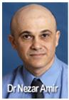
Dr Nezar Amir
Cardiologist
Available at Northern Clinic - Southern Cross North Harbour Campus, 212 Wairau Road, Wairau Valley, Auckland, Tōtara Health, 1 McCrae Way, New Lynn, Auckland
-

Dr Peter Barr
Cardiologist
Available at 1 Gilgit Road, Epsom, Auckland
-

Dr Daniel Chan
Cardiologist
Available at 167 Maunu Road, Maunu, Whangārei 0110
-

Dr Kok Lam Chow
Cardiologist
Available at Ormiston Hospital Specialist Centre & Consulting Suites, 125 Ormiston Road, Flat Bush, Auckland
-
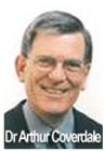
Dr Arthur Coverdale
Cardiologist
Available at 1 Gilgit Road, Epsom, Auckland
-

Dr Gerard Devlin
Cardiologist
Available at 1 Gilgit Road, Epsom, Auckland
-

Prof Rob Doughty
Cardiologist
Available at 1 Gilgit Road, Epsom, Auckland
-

Dr Colin Edwards
Cardiologist
Available at 1 Gilgit Road, Epsom, Auckland, Northern Clinic - Southern Cross North Harbour Campus, 212 Wairau Road, Wairau Valley, Auckland, Tōtara Health, 1 McCrae Way, New Lynn, Auckland
-

Dr Chris Ellis
Cardiologist
Available at 1 Gilgit Road, Epsom, Auckland
-

Dr Shakiya Ershad
Cardiologist
Available at 1 Gilgit Road, Epsom, Auckland
-

Dr Sarah Fitzsimons
Cardiologist
Available at 1 Gilgit Road, Epsom, Auckland
-

Dr Ruvin Gabriel
Cardiologist
Available at 1 Gilgit Road, Epsom, Auckland, Ormiston Hospital Specialist Centre & Consulting Suites, 125 Ormiston Road, Flat Bush, Auckland, Pukekohe Health Centre, 10 West Street, Pukekohe, Auckland
-
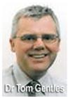
Dr Tom Gentles
Paediatric Cardiologist
Available at 1 Gilgit Road, Epsom, Auckland
-

Dr Ivor Gerber
Cardiologist
Available at 1 Gilgit Road, Epsom, Auckland
-

Dr Wil Harrison
Cardiologist
Available at Ormiston Hospital Specialist Centre & Consulting Suites, 125 Ormiston Road, Flat Bush, Auckland, Pukekohe Health Centre, 10 West Street, Pukekohe, Auckland
-

Dr David Heaven
Cardiologist Electrophysiology
Available at 1 Gilgit Road, Epsom, Auckland
-

Dr Shaw (Anthony) Kueh
Cardiologist
Available at 1 Gilgit Road, Epsom, Auckland
-

Assoc Prof Nigel Lever
Cardiologist
Available at 1 Gilgit Road, Epsom, Auckland
-

Dr Jen-Li Looi
Cardiologist
Available at Ormiston Hospital Specialist Centre & Consulting Suites, 125 Ormiston Road, Flat Bush, Auckland
-

Dr Boris Lowe
Cardiologist
Available at 1 Gilgit Road, Epsom, Auckland
-

Dr Mariana Macedo Lamacie
Cardiologist
Available at 1 Gilgit Road, Epsom, Auckland
-

Dr Andrew Martin
Cardiologist Electrophysiology
Available at 1 Gilgit Road, Epsom, Auckland
-

Dr Bryan Mitchelson
Paediatric Cardiologist
Available at all locations.
-
Dr Ross Nicholson
Paediatric Cardiologist
Available at 1 Gilgit Road, Epsom, Auckland
-

Dr Matthew O'Connor
Cardiologist Electrophysiology
Available at 1 Gilgit Road, Epsom, Auckland
-

Dr Thomas Pasley
Cardiologist
Available at Northern Clinic - Southern Cross North Harbour Campus, 212 Wairau Road, Wairau Valley, Auckland
-

Dr Jeffrey Sebastian
Cardiologist
Available at all locations.
-

Dr Jithendra Somaratne
Cardiologist
Available at 1 Gilgit Road, Epsom, Auckland
-

Dr Fiona Stewart
Cardiologist
Available at 1 Gilgit Road, Epsom, Auckland, eastMED, 188 St Heliers Bay Road, St Heliers, Auckland
-

Prof Ralph Stewart
Cardiologist
Available at 1 Gilgit Road, Epsom, Auckland
-

Dr Michael Stubbs
Cardiologist
Available at 1 Gilgit Road, Epsom, Auckland
-

Dr Timothy Sutton
Cardiologist
Available at 1 Gilgit Road, Epsom, Auckland, Ormiston Hospital Specialist Centre & Consulting Suites, 125 Ormiston Road, Flat Bush, Auckland, Pukekohe Health Centre, 10 West Street, Pukekohe, Auckland
-

Dr Mansi Turaga
Cardiologist
Available at 1 Gilgit Road, Epsom, Auckland, Ormiston Hospital Specialist Centre & Consulting Suites, 125 Ormiston Road, Flat Bush, Auckland
-

Dr Jamie Voss
Cardiologist Electrophysiology
Available at 1 Gilgit Road, Epsom, Auckland
-

Dr Cara Wasywich
Cardiologist
Available at 1 Gilgit Road, Epsom, Auckland, Tōtara Health, 1 McCrae Way, New Lynn, Auckland
-

Dr Jonathon White
Cardiologist
Available at 1 Gilgit Road, Epsom, Auckland
How do I access this service?
Referral, Contact us
Referral Expectations
Your GP will refer you to one of our clinics if they are concerned about your heart and want a specialist opinion.
For all appointments and bookings, please phone (09) 623 1020
Fees and Charges Description
The Heart Group is a Southern Cross, NIB and AIA Affiliated Provider for Cardiac Services. This means that if you have medical insurance cover with these insurance companies, we will complete and submit your claim directly to your medical insurance provider for you.
Charges vary from patient to patient depending on the type of insurance policy held. You are advised of the costs involved at the time of making your appointment.
For all enquiries regarding appointments and charges, please phone (09) 630 1020
Hours
1 Gilgit Road, Epsom, Auckland
| Mon – Thu | 8:00 AM – 8:00 PM |
|---|---|
| Fri – Sat | 8:00 AM – 5:00 PM |
Languages Spoken
Cantonese Chinese, Mandarin Chinese, English
Procedures / Treatments
Cardiology blood tests help doctors check for heart disease, monitor risk factors, and assess how well the heart is working. Some tests check for damage right now (like troponin), while others check your long-term risks (like cholesterol and blood sugar) or stress on the heart (like BNP).
Cardiology blood tests help doctors check for heart disease, monitor risk factors, and assess how well the heart is working. Some tests check for damage right now (like troponin), while others check your long-term risks (like cholesterol and blood sugar) or stress on the heart (like BNP).
Cardiology blood tests help doctors check for heart disease, monitor risk factors, and assess how well the heart is working. Some tests check for damage right now (like troponin), while others check your long-term risks (like cholesterol and blood sugar) or stress on the heart (like BNP).
Heart rhythm refers to the electrical source that is driving the heart rate and whether or not it is regular or irregular. Heart rhythm can be affected by a number of conditions. Some common terms Sinus rhythm is the normal rhythm Arrhythmia means abnormal rhythm Fibrillation means irregular rhythm or quivering of one part of the heart Bradycardia means slow heart rate Tachycardia means fast heart rate Paroxysmal means the arrhythmia comes and goes Tachycardia The most common form of this is atrial fibrillation. This is where the heart rhythm is irregular and often too fast. Symptoms include fatigue, palpitations (where you are aware of your heart racing or pounding), dizziness and breathlessness. Other tachycardias include supraventricular tachycardia (SVT) or ventricular tachycardia (VT). These have similar symptoms as atrial fibrillation but can also cause you to lose consciousness (faint). Bradycardia The most common form of this is called heart block. This is because messages from the electrical generator of the heart don't get through efficiently to the rest of the heart and hence it goes very slowly or can pause. Symptoms of the heart going too slowly include feeling tired, breathless or fainting. Tests Tests to diagnose what sort of arrhythmia you have include an electrocardiogram (ECG) and an ambulatory ECG (Holter monitor or Event recorder). Treatment Most treatments for tachycardias consist of medication to stop the abnormal rhythm or make it slower if and when it occurs. Atrial fibrillation, if you have other problems, can increase your risk of stroke so blood-thinning medication is often used as well. If you have bradycardia, you may be referred to the surgeons for a pacemaker. This is a small operation where a battery powered device is placed under the skin with wires that lead to your heart and provide it with electrical stimulation to prevent it from going too slowly. You can't feel it doing this but will be aware of a small flat lump under your skin just below your collar bone.
Heart rhythm refers to the electrical source that is driving the heart rate and whether or not it is regular or irregular. Heart rhythm can be affected by a number of conditions. Some common terms Sinus rhythm is the normal rhythm Arrhythmia means abnormal rhythm Fibrillation means irregular rhythm or quivering of one part of the heart Bradycardia means slow heart rate Tachycardia means fast heart rate Paroxysmal means the arrhythmia comes and goes Tachycardia The most common form of this is atrial fibrillation. This is where the heart rhythm is irregular and often too fast. Symptoms include fatigue, palpitations (where you are aware of your heart racing or pounding), dizziness and breathlessness. Other tachycardias include supraventricular tachycardia (SVT) or ventricular tachycardia (VT). These have similar symptoms as atrial fibrillation but can also cause you to lose consciousness (faint). Bradycardia The most common form of this is called heart block. This is because messages from the electrical generator of the heart don't get through efficiently to the rest of the heart and hence it goes very slowly or can pause. Symptoms of the heart going too slowly include feeling tired, breathless or fainting. Tests Tests to diagnose what sort of arrhythmia you have include an electrocardiogram (ECG) and an ambulatory ECG (Holter monitor or Event recorder). Treatment Most treatments for tachycardias consist of medication to stop the abnormal rhythm or make it slower if and when it occurs. Atrial fibrillation, if you have other problems, can increase your risk of stroke so blood-thinning medication is often used as well. If you have bradycardia, you may be referred to the surgeons for a pacemaker. This is a small operation where a battery powered device is placed under the skin with wires that lead to your heart and provide it with electrical stimulation to prevent it from going too slowly. You can't feel it doing this but will be aware of a small flat lump under your skin just below your collar bone.
Heart rhythm refers to the electrical source that is driving the heart rate and whether or not it is regular or irregular. Heart rhythm can be affected by a number of conditions.
Some common terms
- Sinus rhythm is the normal rhythm
- Arrhythmia means abnormal rhythm
- Fibrillation means irregular rhythm or quivering of one part of the heart
- Bradycardia means slow heart rate
- Tachycardia means fast heart rate
- Paroxysmal means the arrhythmia comes and goes
Tachycardia
The most common form of this is atrial fibrillation. This is where the heart rhythm is irregular and often too fast. Symptoms include fatigue, palpitations (where you are aware of your heart racing or
pounding), dizziness and breathlessness.
Other tachycardias include supraventricular tachycardia (SVT) or ventricular tachycardia (VT). These have similar symptoms as atrial fibrillation but can also cause you to lose consciousness (faint).
Bradycardia
The most common form of this is called heart block. This is because messages from the electrical generator of the heart don't get through efficiently to the rest of the heart and hence it goes very slowly or can pause. Symptoms of the heart going too slowly include feeling tired, breathless or fainting.
Tests
Tests to diagnose what sort of arrhythmia you have include an electrocardiogram (ECG) and an ambulatory ECG (Holter monitor or Event recorder).
Treatment
Most treatments for tachycardias consist of medication to stop the abnormal rhythm or make it slower if and when it occurs. Atrial fibrillation, if you have other problems, can increase your risk of stroke so blood-thinning medication is often used as well.
If you have bradycardia, you may be referred to the surgeons for a pacemaker. This is a small operation where a battery powered device is placed under the skin with wires that lead to your heart and provide it with electrical stimulation to prevent it from going too slowly. You can't feel it doing this but will be aware of a small flat lump under your skin just below your collar bone.
Cardiovascular disease is a general term for any condition that affects the heart or blood vessels. Common types of cardiovascular disease include: Coronary artery disease – narrowing or blockage of the arteries that supply the heart, which can lead to chest pain or heart attack. Heart failure – when the heart can’t pump blood effectively. Arrhythmias – abnormal heart rhythms. Stroke – damage to the brain caused by blocked or burst blood vessels. Peripheral artery disease – narrowing of arteries in the legs or arms. Heart valve problems – issues with valves that control blood flow through the heart. Risk factors: High blood pressure High cholesterol Smoking Diabetes Obesity Family history of heart disease Are older (your risk increases as you get older)
Cardiovascular disease is a general term for any condition that affects the heart or blood vessels. Common types of cardiovascular disease include: Coronary artery disease – narrowing or blockage of the arteries that supply the heart, which can lead to chest pain or heart attack. Heart failure – when the heart can’t pump blood effectively. Arrhythmias – abnormal heart rhythms. Stroke – damage to the brain caused by blocked or burst blood vessels. Peripheral artery disease – narrowing of arteries in the legs or arms. Heart valve problems – issues with valves that control blood flow through the heart. Risk factors: High blood pressure High cholesterol Smoking Diabetes Obesity Family history of heart disease Are older (your risk increases as you get older)
Cardiovascular disease is a general term for any condition that affects the heart or blood vessels.
Common types of cardiovascular disease include:
- Coronary artery disease – narrowing or blockage of the arteries that supply the heart, which can lead to chest pain or heart attack.
- Heart failure – when the heart can’t pump blood effectively.
- Arrhythmias – abnormal heart rhythms.
- Stroke – damage to the brain caused by blocked or burst blood vessels.
- Peripheral artery disease – narrowing of arteries in the legs or arms.
- Heart valve problems – issues with valves that control blood flow through the heart.
Risk factors:
- High blood pressure
- High cholesterol
- Smoking
- Diabetes
- Obesity
- Family history of heart disease
- Are older (your risk increases as you get older)
This test is performed by a cardiologist in a sterile operating theatre environment. Most people will need to have routine tests before the procedure. These tests may require separate appointments and are usually planned the day before or the day of the procedure. You will be asked not to eat or drink after midnight the evening before the procedure. You are not given a general anaesthetic but may have some medication to relax you if needed. Local anaesthetic is put into an area of skin to the side of your groin or in your arm. A needle and then tube are fed into an artery here and advanced through the blood vessels to the heart. Dye is then injected so that the heart and blood vessels can be seen on X-ray. X-rays and measurements are then taken giving the doctors information about the state of your heart and the exact nature of any narrowed blood vessels. This allows them to plan the best form of treatment to prevent heart attacks and control any symptoms you may have. After the procedure you will have to lay flat for several hours to prevent bleeding.
This test is performed by a cardiologist in a sterile operating theatre environment. Most people will need to have routine tests before the procedure. These tests may require separate appointments and are usually planned the day before or the day of the procedure. You will be asked not to eat or drink after midnight the evening before the procedure. You are not given a general anaesthetic but may have some medication to relax you if needed. Local anaesthetic is put into an area of skin to the side of your groin or in your arm. A needle and then tube are fed into an artery here and advanced through the blood vessels to the heart. Dye is then injected so that the heart and blood vessels can be seen on X-ray. X-rays and measurements are then taken giving the doctors information about the state of your heart and the exact nature of any narrowed blood vessels. This allows them to plan the best form of treatment to prevent heart attacks and control any symptoms you may have. After the procedure you will have to lay flat for several hours to prevent bleeding.
This test is performed by a cardiologist in a sterile operating theatre environment.
Most people will need to have routine tests before the procedure. These tests may require separate appointments and are usually planned the day before or the day of the procedure. You will be asked not to eat or drink after midnight the evening before the procedure.
You are not given a general anaesthetic but may have some medication to relax you if needed. Local anaesthetic is put into an area of skin to the side of your groin or in your arm. A needle and then tube are fed into an artery here and advanced through the blood vessels to the heart. Dye is then injected so that the heart and blood vessels can be seen on X-ray. X-rays and measurements are then taken giving the doctors information about the state of your heart and the exact nature of any narrowed blood vessels. This allows them to plan the best form of treatment to prevent heart attacks and control any symptoms you may have.
After the procedure you will have to lay flat for several hours to prevent bleeding.
Echocardiography (or cardiac ultrasound) is a test that uses high frequency sound waves to generate pictures of your heart. During the test, you generally lie on your back, gel is applied to your skin and a technician then moves the small, plastic transducer over your chest. The test is painless and can take from 10 minutes to an hour. The machine then develops images of your heart which are seen on a monitor. This is referred to as an echocardiogram. Echocardiography can help in the diagnosis of many heart problems including cardiovascular disease, previous heart attacks, valve disorders, weakened heart muscle, holes between heart chambers, fluid around the heart (pericardial effusion). If doctors are looking for evidence of coronary artery disease, they may perform variations of this test which include: Exercise echocardiography - compares how your heart works when stressed by exercise versus when it is at rest. The ultrasound is conducted before you exercise and immediately after you stop. Either a stationary bicycle or standard treadmill is used. Dobutamine stress echocardiography - if you’re unable to exercise for the above test, you might be given medication to simulate the effects of exercise. During this test, an echocardiogram initially is performed when you’re at rest. Then dobutamine is given to you via a needle into a vein in your arm. Its effect is to make your heart work harder and faster just like with exercise. After it has taken effect, the echocardiogram is repeated. The effect wears off very quickly.
Echocardiography (or cardiac ultrasound) is a test that uses high frequency sound waves to generate pictures of your heart. During the test, you generally lie on your back, gel is applied to your skin and a technician then moves the small, plastic transducer over your chest. The test is painless and can take from 10 minutes to an hour. The machine then develops images of your heart which are seen on a monitor. This is referred to as an echocardiogram. Echocardiography can help in the diagnosis of many heart problems including cardiovascular disease, previous heart attacks, valve disorders, weakened heart muscle, holes between heart chambers, fluid around the heart (pericardial effusion). If doctors are looking for evidence of coronary artery disease, they may perform variations of this test which include: Exercise echocardiography - compares how your heart works when stressed by exercise versus when it is at rest. The ultrasound is conducted before you exercise and immediately after you stop. Either a stationary bicycle or standard treadmill is used. Dobutamine stress echocardiography - if you’re unable to exercise for the above test, you might be given medication to simulate the effects of exercise. During this test, an echocardiogram initially is performed when you’re at rest. Then dobutamine is given to you via a needle into a vein in your arm. Its effect is to make your heart work harder and faster just like with exercise. After it has taken effect, the echocardiogram is repeated. The effect wears off very quickly.
Echocardiography (or cardiac ultrasound) is a test that uses high frequency sound waves to generate pictures of your heart. During the test, you generally lie on your back, gel is applied to your skin and a technician then moves the small, plastic transducer over your chest. The test is painless and can take from 10 minutes to an hour.
The machine then develops images of your heart which are seen on a monitor. This is referred to as an echocardiogram.
Echocardiography can help in the diagnosis of many heart problems including cardiovascular disease, previous heart attacks, valve disorders, weakened heart muscle, holes between heart chambers, fluid around the heart (pericardial effusion).
If doctors are looking for evidence of coronary artery disease, they may perform variations of this test which include:
- Exercise echocardiography - compares how your heart works when stressed by exercise versus when it is at rest. The ultrasound is conducted before you exercise and immediately after you stop. Either a stationary bicycle or standard treadmill is used.
- Dobutamine stress echocardiography - if you’re unable to exercise for the above test, you might be given medication to simulate the effects of exercise. During this test, an echocardiogram initially is performed when you’re at rest. Then dobutamine is given to you via a needle into a vein in your arm. Its effect is to make your heart work harder and faster just like with exercise. After it has taken effect, the echocardiogram is repeated. The effect wears off very quickly.
An ECG is a recording of your heart's electrical activity. Electrode patches are attached to your skin to measure the electrical impulses given off by your heart. The result is a trace that can be read by a doctor. It can give information of previous heart attacks or problems with the heart rhythm. Ambulatory ECG - this can be performed with a Holter monitor which monitors your heart for rhythm abnormalities during normal activity for an uninterrupted 24-hour period. During the test, electrodes attached to your chest are connected to a portable recorder - about the size of a paperback book - that's attached to your belt or hung from a shoulder strap. Another form of ambulatory ECG test is an Event recorder which covers 1-2 weeks. You wear a monitor (much smaller than a Holter monitor) and if you have any symptoms, such as dizziness, you press a button on a recording device which saves the recording of your heart rhythm made in the minutes leading up to and during your symptoms. Because you can wear this for a longer period of time it has a higher rate of catching your abnormal rhythm.
An ECG is a recording of your heart's electrical activity. Electrode patches are attached to your skin to measure the electrical impulses given off by your heart. The result is a trace that can be read by a doctor. It can give information of previous heart attacks or problems with the heart rhythm. Ambulatory ECG - this can be performed with a Holter monitor which monitors your heart for rhythm abnormalities during normal activity for an uninterrupted 24-hour period. During the test, electrodes attached to your chest are connected to a portable recorder - about the size of a paperback book - that's attached to your belt or hung from a shoulder strap. Another form of ambulatory ECG test is an Event recorder which covers 1-2 weeks. You wear a monitor (much smaller than a Holter monitor) and if you have any symptoms, such as dizziness, you press a button on a recording device which saves the recording of your heart rhythm made in the minutes leading up to and during your symptoms. Because you can wear this for a longer period of time it has a higher rate of catching your abnormal rhythm.
An ECG is a recording of your heart's electrical activity. Electrode patches are attached to your skin to measure the electrical impulses given off by your heart. The result is a trace that can be read by a doctor. It can give information of previous heart attacks or problems with the heart rhythm.
Ambulatory ECG - this can be performed with a Holter monitor which monitors your heart for rhythm abnormalities during normal activity for an uninterrupted 24-hour period. During the test, electrodes attached to your chest are connected to a portable recorder - about the size of a paperback book - that's attached to your belt or hung from a shoulder strap.
Another form of ambulatory ECG test is an Event recorder which covers 1-2 weeks. You wear a monitor (much smaller than a Holter monitor) and if you have any symptoms, such as dizziness, you press a button on a recording device which saves the recording of your heart rhythm made in the minutes leading up to and during your symptoms. Because you can wear this for a longer period of time it has a higher rate of catching your abnormal rhythm.
An ECG done when you are resting may be normal even when you have cardiovascular disease. During an exercise ECG the heart is made to work harder so that if there is any narrowing of the blood vessels resulting in poor blood supply it is more likely to be picked up on the tracing as your heart goes faster. For this test you have to work harder which involves walking on a treadmill while your heart is monitored. The treadmill gets faster with time but you can stop at anytime. This test is supervised and interpreted by a doctor as you go. This test is used to see if you have any evidence of cardiovascular disease and can give the doctor some idea as to how severe it might be so as to direct further tests and possible treatment.
An ECG done when you are resting may be normal even when you have cardiovascular disease. During an exercise ECG the heart is made to work harder so that if there is any narrowing of the blood vessels resulting in poor blood supply it is more likely to be picked up on the tracing as your heart goes faster. For this test you have to work harder which involves walking on a treadmill while your heart is monitored. The treadmill gets faster with time but you can stop at anytime. This test is supervised and interpreted by a doctor as you go. This test is used to see if you have any evidence of cardiovascular disease and can give the doctor some idea as to how severe it might be so as to direct further tests and possible treatment.
An ECG done when you are resting may be normal even when you have cardiovascular disease. During an exercise ECG the heart is made to work harder so that if there is any narrowing of the blood vessels resulting in poor blood supply it is more likely to be picked up on the tracing as your heart goes faster. For this test you have to work harder which involves walking on a treadmill while your heart is monitored. The treadmill gets faster with time but you can stop at anytime. This test is supervised and interpreted by a doctor as you go. This test is used to see if you have any evidence of cardiovascular disease and can give the doctor some idea as to how severe it might be so as to direct further tests and possible treatment.
Heart failure refers to the heart failing to pump efficiently. There are many diseases that cause this including cardiovascular disease, high blood pressure, viral infections, alcohol, and diseases affecting the valves of the heart. When the heart is inefficient a number of symptoms occur depending on the cause and severity of the condition. The main symptoms are tiredness, breathlessness on exertion or lying flat, and ankle swelling. Doctors often refer to oedema, which means fluid retention usually in your feet or lungs as a result of the heart not pumping efficiently. Tests looking for possible causes of heart failure include: Chest x-ray, Electrocardiogram (ECG), Echocardiogram (Cardiac ultrasound), Angiogram. You are likely to be given several medications over time, started and monitored by your cardiologist and GP. These include medication to control the amount of fluid that builds up (diuretics), medication to protect your heart and slow it down as well as to thin your blood. You will often be referred to a dietitian or given advice about restricting the amount of fluid and salt you take as this can contribute to symptoms.
Heart failure refers to the heart failing to pump efficiently. There are many diseases that cause this including cardiovascular disease, high blood pressure, viral infections, alcohol, and diseases affecting the valves of the heart. When the heart is inefficient a number of symptoms occur depending on the cause and severity of the condition. The main symptoms are tiredness, breathlessness on exertion or lying flat, and ankle swelling. Doctors often refer to oedema, which means fluid retention usually in your feet or lungs as a result of the heart not pumping efficiently. Tests looking for possible causes of heart failure include: Chest x-ray, Electrocardiogram (ECG), Echocardiogram (Cardiac ultrasound), Angiogram. You are likely to be given several medications over time, started and monitored by your cardiologist and GP. These include medication to control the amount of fluid that builds up (diuretics), medication to protect your heart and slow it down as well as to thin your blood. You will often be referred to a dietitian or given advice about restricting the amount of fluid and salt you take as this can contribute to symptoms.
Heart failure refers to the heart failing to pump efficiently. There are many diseases that cause this including cardiovascular disease, high blood pressure, viral infections, alcohol, and diseases affecting the valves of the heart. When the heart is inefficient a number of symptoms occur depending on the cause and severity of the condition. The main symptoms are tiredness, breathlessness on exertion or lying flat, and ankle swelling. Doctors often refer to oedema, which means fluid retention usually in your feet or lungs as a result of the heart not pumping efficiently.
Tests looking for possible causes of heart failure include: Chest x-ray, Electrocardiogram (ECG), Echocardiogram (Cardiac ultrasound), Angiogram.
You are likely to be given several medications over time, started and monitored by your cardiologist and GP. These include medication to control the amount of fluid that builds up (diuretics), medication to protect your heart and slow it down as well as to thin your blood. You will often be referred to a dietitian or given advice about restricting the amount of fluid and salt you take as this can contribute to symptoms.
Your heart consists of four chambers that receive and send blood to the lungs and body. Disorders affecting valves can either cause stenosis (a narrowing) or regurgitation (leakage after the valve has closed). Depending on what valve is involved and how severe the damage is it may result in symptoms of heart failure, as it makes the heart pump inefficiently. Suspicion of a heart valve problem is usually picked up by your doctor when they listen to your heart and hear a murmur. A murmur is heard with the stethoscope and is turbulence of blood flow that occurs through a narrowed or leaky valve. Not all heart murmurs mean serious problems but are best investigated further. The echocardiogram is the main test to diagnose what valve is involved and how severe it is. Treatment depends on the type and severity of the valve lesion. You may simply be monitored over years to see if anything changes. Some conditions require medication to thin the blood or treat any complicating heart problems. You may be referred to a heart surgeon for consideration of a valve replacement or dilatation of a narrowed valve.
Your heart consists of four chambers that receive and send blood to the lungs and body. Disorders affecting valves can either cause stenosis (a narrowing) or regurgitation (leakage after the valve has closed). Depending on what valve is involved and how severe the damage is it may result in symptoms of heart failure, as it makes the heart pump inefficiently. Suspicion of a heart valve problem is usually picked up by your doctor when they listen to your heart and hear a murmur. A murmur is heard with the stethoscope and is turbulence of blood flow that occurs through a narrowed or leaky valve. Not all heart murmurs mean serious problems but are best investigated further. The echocardiogram is the main test to diagnose what valve is involved and how severe it is. Treatment depends on the type and severity of the valve lesion. You may simply be monitored over years to see if anything changes. Some conditions require medication to thin the blood or treat any complicating heart problems. You may be referred to a heart surgeon for consideration of a valve replacement or dilatation of a narrowed valve.
Your heart consists of four chambers that receive and send blood to the lungs and body.
Disorders affecting valves can either cause stenosis (a narrowing) or regurgitation (leakage after the valve has closed). Depending on what valve is involved and how severe the damage is it may result in symptoms of heart failure, as it makes the heart pump inefficiently.
Suspicion of a heart valve problem is usually picked up by your doctor when they listen to your heart and hear a murmur. A murmur is heard with the stethoscope and is turbulence of blood flow that occurs through a narrowed or leaky valve. Not all heart murmurs mean serious problems but are best investigated further.
The echocardiogram is the main test to diagnose what valve is involved and how severe it is.
Treatment depends on the type and severity of the valve lesion. You may simply be monitored over years to see if anything changes. Some conditions require medication to thin the blood or treat any complicating heart problems. You may be referred to a heart surgeon for consideration of a valve replacement or dilatation of a narrowed valve.
Parking
Patient parking is provided at all locations.
- 1 Gilgit Road - entry from Gilgit Road only
- New Lynn: patient parking is available on Level 1, Totara Health Services, 1 McCrae Way.
- Ormiston Hospital - please note paid parking at this location.
Pharmacy
Other
| SERVICE & HOURS | |
|
CENTRAL |
Monday to Friday including evenings and Saturdays: Consulting & Echocardiography Phone 09 623 1020 |
| NORTH | Southern Cross Hospital, North Harbour Weekday Clinics Ph 09 623 1020 |
| EAST | EastMed, St Heliers Weekday Clinics Ph 09 623 1020 |
| WEST | Totara Health Services, New Lynn Weekday Clinics Ph 09 623 1020 |
| SOUTH |
Ormiston Hospital, Botany |
| SOUTH |
Pukekohe Family Health Centre Weekday Clinics Ph 09 623 1020 |
Website

Contact Details
1 Gilgit Road, Epsom, Auckland
Central Auckland
-
Phone
(09) 623 1020
-
Fax
(09) 623 1030
Healthlink EDI
akhearta
Email
Website
1 Gilgit Road
Epsom
Auckland 1023
Street Address
1 Gilgit Road
Epsom
Auckland 1023
Postal Address
PO Box 99500
Newmarket
Auckland 1149
Northern Clinic - Southern Cross North Harbour Campus, 212 Wairau Road, Wairau Valley, Auckland
North Auckland
-
Phone
(09) 623 1020
-
Fax
(09) 623 1030
Healthlink EDI
akhearta
Email
Website
Ormiston Hospital Specialist Centre & Consulting Suites, 125 Ormiston Road, Flat Bush, Auckland
South Auckland
Healthlink EDI
akhearta
Email
Website
Tōtara Health, 1 McCrae Way, New Lynn, Auckland
West Auckland
-
Phone
(09) 623 1020
-
Fax
(09) 623 1030
Healthlink EDI
akhearta
Email
Website
eastMED, 188 St Heliers Bay Road, St Heliers, Auckland
Central Auckland
-
Phone
(09) 623 1020
-
Fax
(09) 623 1030
Healthlink EDI
akhearta
Email
Website
Pukekohe Health Centre, 10 West Street, Pukekohe, Auckland
South Auckland
-
Phone
(09) 623 1020
-
Fax
(09) 623 1030
Healthlink EDI
akhearta
Email
Website
167 Maunu Road, Maunu, Whangārei 0110
Northland
-
Phone
(09) 623 1020
-
Fax
(09) 623 1030
Healthlink EDI
akhearta
Email
Website
Was this page helpful?
This page was last updated at 11:27AM on March 19, 2026. This information is reviewed and edited by The Heart Group.