

Central Auckland, East Auckland, North Auckland, South Auckland, West Auckland > Private Hospitals & Specialists >
The Specialists Takapuna - General, Bariatric, Breast, Skin Cancer & Thyroid Surgery
Private Surgical Service, General Surgery, Breast
Description
We are a new Surgical Centre offering state-of-the-art facilities in the heart of Takapuna.
Consultants
-

Dr Katherine Gale
Oncoplastic Breast Surgeon
-
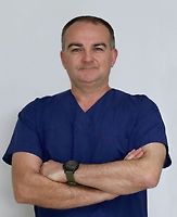
Mr Richard Martin
General Surgeon
-

Mr Jason Robertson
Upper GI and Bariatic Surgeon
Procedures / Treatments
Shave Biopsy: the top layers of skin in the area being investigated are shaved off with a scalpel (surgical knife) for investigation under a microscope. Punch Biopsy: a small cylindrical core of tissue is taken from the area being investigated for examination under a microscope. Excision Biopsy: all of the lesion or area being investigated is cut out with a scalpel for examination under a microscope. Incision Biopsy: part of the lesion is cut out with a scalpel for examination under a microscope.
Shave Biopsy: the top layers of skin in the area being investigated are shaved off with a scalpel (surgical knife) for investigation under a microscope. Punch Biopsy: a small cylindrical core of tissue is taken from the area being investigated for examination under a microscope. Excision Biopsy: all of the lesion or area being investigated is cut out with a scalpel for examination under a microscope. Incision Biopsy: part of the lesion is cut out with a scalpel for examination under a microscope.
Shave Biopsy: the top layers of skin in the area being investigated are shaved off with a scalpel (surgical knife) for investigation under a microscope.
Punch Biopsy: a small cylindrical core of tissue is taken from the area being investigated for examination under a microscope.
Excision Biopsy: all of the lesion or area being investigated is cut out with a scalpel for examination under a microscope.
Incision Biopsy: part of the lesion is cut out with a scalpel for examination under a microscope.
Skin lesions such as cysts and tumours are removed by cutting around and under them with a scalpel.
Skin lesions such as cysts and tumours are removed by cutting around and under them with a scalpel.
Skin lesions such as cysts and tumours are removed by cutting around and under them with a scalpel.
A hernia exists where part of the abdominal wall is weakened, and the contents of the abdomen push through to the outside. This is most commonly seen in the groin area but can occur in other places. Surgical treatment is usually quite straightforward and involves returning the abdominal contents to the inside and then reinforcing the abdominal wall in some way. Hiatus Hernia: Laparoscopic: several small incisions (cuts) are made in the abdomen (stomach) and a narrow tube with a tiny camera attached (laparoscope) is inserted. Small instruments are inserted through the other cuts, allowing the surgeon to push the hernia (part of the stomach and lower oesophagus that is bulging into the chest) back into position in the abdominal cavity. The hiatus (opening) in the diaphragm (a sheet of muscle between the chest and stomach) is tightened and the stomach is stitched into place. Open: an abdominal incision is made over the hernia and the hernia is pushed back into position in the abdominal cavity. The hiatus (opening in the diaphragm) is tightened and the stomach is stitched into place. Fundoplication: during the above procedures, the top part of the stomach (fundus) may be secured in position by wrapping it around the oesophagus. Inguinal Hernia: Laparoscopic: several small incisions are made in the abdomen and a narrow tube with a tiny camera attached (laparoscope) is inserted. Small instruments are inserted through the other cuts, allowing the surgeon to push the hernia (part of the intestine that is bulging through the abdominal wall) back into its original position. The weakness in the abdominal wall is repaired. Open: an abdominal incision is made and the hernia is pushed back into position. The weakness in the abdominal wall is repaired. Umbilical Hernia: An incision is made underneath the navel (tummy button) and the hernia (part of the intestine that is bulging through the abdominal wall) is pushed back into the abdominal cavity. The weakness in the abdominal wall is repaired. Incisional Hernia: Laparoscopic: several small incisions are made in the abdomen and a narrow tube with a tiny camera attached (laparoscope) is inserted. Small instruments are inserted through the other cuts, allowing the surgeon to push the hernia (part of the intestine that is bulging through the abdominal wall) back into its original position. Open: an abdominal incision is made and the hernia is pushed back into position.
A hernia exists where part of the abdominal wall is weakened, and the contents of the abdomen push through to the outside. This is most commonly seen in the groin area but can occur in other places. Surgical treatment is usually quite straightforward and involves returning the abdominal contents to the inside and then reinforcing the abdominal wall in some way. Hiatus Hernia: Laparoscopic: several small incisions (cuts) are made in the abdomen (stomach) and a narrow tube with a tiny camera attached (laparoscope) is inserted. Small instruments are inserted through the other cuts, allowing the surgeon to push the hernia (part of the stomach and lower oesophagus that is bulging into the chest) back into position in the abdominal cavity. The hiatus (opening) in the diaphragm (a sheet of muscle between the chest and stomach) is tightened and the stomach is stitched into place. Open: an abdominal incision is made over the hernia and the hernia is pushed back into position in the abdominal cavity. The hiatus (opening in the diaphragm) is tightened and the stomach is stitched into place. Fundoplication: during the above procedures, the top part of the stomach (fundus) may be secured in position by wrapping it around the oesophagus. Inguinal Hernia: Laparoscopic: several small incisions are made in the abdomen and a narrow tube with a tiny camera attached (laparoscope) is inserted. Small instruments are inserted through the other cuts, allowing the surgeon to push the hernia (part of the intestine that is bulging through the abdominal wall) back into its original position. The weakness in the abdominal wall is repaired. Open: an abdominal incision is made and the hernia is pushed back into position. The weakness in the abdominal wall is repaired. Umbilical Hernia: An incision is made underneath the navel (tummy button) and the hernia (part of the intestine that is bulging through the abdominal wall) is pushed back into the abdominal cavity. The weakness in the abdominal wall is repaired. Incisional Hernia: Laparoscopic: several small incisions are made in the abdomen and a narrow tube with a tiny camera attached (laparoscope) is inserted. Small instruments are inserted through the other cuts, allowing the surgeon to push the hernia (part of the intestine that is bulging through the abdominal wall) back into its original position. Open: an abdominal incision is made and the hernia is pushed back into position.
A hernia exists where part of the abdominal wall is weakened, and the contents of the abdomen push through to the outside. This is most commonly seen in the groin area but can occur in other places. Surgical treatment is usually quite straightforward and involves returning the abdominal contents to the inside and then reinforcing the abdominal wall in some way.
Hiatus Hernia:
Laparoscopic: several small incisions (cuts) are made in the abdomen (stomach) and a narrow tube with a tiny camera attached (laparoscope) is inserted. Small instruments are inserted through the other cuts, allowing the surgeon to push the hernia (part of the stomach and lower oesophagus that is bulging into the chest) back into position in the abdominal cavity. The hiatus (opening) in the diaphragm (a sheet of muscle between the chest and stomach) is tightened and the stomach is stitched into place.
Open: an abdominal incision is made over the hernia and the hernia is pushed back into position in the abdominal cavity. The hiatus (opening in the diaphragm) is tightened and the stomach is stitched into place.
Fundoplication: during the above procedures, the top part of the stomach (fundus) may be secured in position by wrapping it around the oesophagus.
Inguinal Hernia:
Laparoscopic: several small incisions are made in the abdomen and a narrow tube with a tiny camera attached (laparoscope) is inserted. Small instruments are inserted through the other cuts, allowing the surgeon to push the hernia (part of the intestine that is bulging through the abdominal wall) back into its original position. The weakness in the abdominal wall is repaired.
Open: an abdominal incision is made and the hernia is pushed back into position. The weakness in the abdominal wall is repaired.
Umbilical Hernia:
An incision is made underneath the navel (tummy button) and the hernia (part of the intestine that is bulging through the abdominal wall) is pushed back into the abdominal cavity. The weakness in the abdominal wall is repaired.
Incisional Hernia:
Laparoscopic: several small incisions are made in the abdomen and a narrow tube with a tiny camera attached (laparoscope) is inserted. Small instruments are inserted through the other cuts, allowing the surgeon to push the hernia (part of the intestine that is bulging through the abdominal wall) back into its original position.
Open: an abdominal incision is made and the hernia is pushed back into position.
Laparoscopic: several small incisions (cuts) are made in the lower right abdomen (stomach) and a narrow tube with a tiny camera attached (laparoscope) in inserted. This allows the surgeon a view of the appendix and, by inserting small surgical instruments through the other cuts, the appendix can be removed. Open: an incision is made in the lower right abdomen and the appendix removed.
Laparoscopic: several small incisions (cuts) are made in the lower right abdomen (stomach) and a narrow tube with a tiny camera attached (laparoscope) in inserted. This allows the surgeon a view of the appendix and, by inserting small surgical instruments through the other cuts, the appendix can be removed. Open: an incision is made in the lower right abdomen and the appendix removed.
Laparoscopic: several small incisions (cuts) are made in the lower right abdomen (stomach) and a narrow tube with a tiny camera attached (laparoscope) in inserted. This allows the surgeon a view of the appendix and, by inserting small surgical instruments through the other cuts, the appendix can be removed.
Open: an incision is made in the lower right abdomen and the appendix removed.
Gallstones are formed if the gallbladder is not working properly, and the standard treatment is to remove the gallbladder (cholecystectomy). This procedure is usually performed using a laparoscopic (keyhole) approach. Laparoscopic: several small incisions (cuts) are made in the abdomen (stomach) and a narrow tube with a tiny camera attached (laparoscope) is inserted. This allows the surgeon a view of the gallbladder and, by inserting small surgical instruments through the other cuts, the gallbladder can be removed. Open: an abdominal incision is made and the gallbladder removed.
Gallstones are formed if the gallbladder is not working properly, and the standard treatment is to remove the gallbladder (cholecystectomy). This procedure is usually performed using a laparoscopic (keyhole) approach. Laparoscopic: several small incisions (cuts) are made in the abdomen (stomach) and a narrow tube with a tiny camera attached (laparoscope) is inserted. This allows the surgeon a view of the gallbladder and, by inserting small surgical instruments through the other cuts, the gallbladder can be removed. Open: an abdominal incision is made and the gallbladder removed.
Gallstones are formed if the gallbladder is not working properly, and the standard treatment is to remove the gallbladder (cholecystectomy). This procedure is usually performed using a laparoscopic (keyhole) approach.
Laparoscopic: several small incisions (cuts) are made in the abdomen (stomach) and a narrow tube with a tiny camera attached (laparoscope) is inserted. This allows the surgeon a view of the gallbladder and, by inserting small surgical instruments through the other cuts, the gallbladder can be removed.
Open: an abdominal incision is made and the gallbladder removed.
Partial: the diseased part of the stomach is removed and the remaining section is reattached to the oesophagus (food pipe) or small intestine. Total: all of the stomach is removed and the oesophagus is attached directly to the small intestine.
Partial: the diseased part of the stomach is removed and the remaining section is reattached to the oesophagus (food pipe) or small intestine. Total: all of the stomach is removed and the oesophagus is attached directly to the small intestine.
Partial: the diseased part of the stomach is removed and the remaining section is reattached to the oesophagus (food pipe) or small intestine.
Total: all of the stomach is removed and the oesophagus is attached directly to the small intestine.
Gastroscopy allows examination of the upper part of your digestive tract i.e. oesophagus (food pipe), stomach and duodenum (top section of the small intestine), by passing a gastroscope (long, flexible tube with a camera on the end) through your mouth and down your digestive tract. Images from the camera are displayed on a television monitor. Sometimes a small tissue sample (biopsy) will need to be taken during the procedure for later examination at a laboratory. Gastroscopy may be used to diagnose peptic ulcers, tumours, gastritis etc. Complications from this procedure are very rare but can occur. They include: bleeding if a biopsy is performed; allergic reaction to the sedative or throat spray; perforation (tearing) of the stomach with the instrument (this is a serious but extremely rare complication). What to expect All endoscopic procedures are viewed as a surgical procedure and generally the same preparation will apply. You will not be able to eat or drink anything for 6 hours before your gastroscopy. When you are ready for the procedure, the back of your throat will be sprayed with anaesthetic. You will also be offered medication (a sedative) to make you go into a light sleep. This will be given by an injection into a vein in your arm or hand. The gastroscopy will take approximately 15 minutes, but you will probably sleep for another 30 minutes. You will spend some time in a recovery unit (probably 1-2 hours) to sleep off the sedative and to allow staff to monitor you (take blood pressure readings etc). Because you have been sedated (given medication to make you sleep) it is important that you arrange for someone else to drive you home. If biopsies are taken for examination, your GP will be sent the results within 2-3 weeks.
Gastroscopy allows examination of the upper part of your digestive tract i.e. oesophagus (food pipe), stomach and duodenum (top section of the small intestine), by passing a gastroscope (long, flexible tube with a camera on the end) through your mouth and down your digestive tract. Images from the camera are displayed on a television monitor. Sometimes a small tissue sample (biopsy) will need to be taken during the procedure for later examination at a laboratory. Gastroscopy may be used to diagnose peptic ulcers, tumours, gastritis etc. Complications from this procedure are very rare but can occur. They include: bleeding if a biopsy is performed; allergic reaction to the sedative or throat spray; perforation (tearing) of the stomach with the instrument (this is a serious but extremely rare complication). What to expect All endoscopic procedures are viewed as a surgical procedure and generally the same preparation will apply. You will not be able to eat or drink anything for 6 hours before your gastroscopy. When you are ready for the procedure, the back of your throat will be sprayed with anaesthetic. You will also be offered medication (a sedative) to make you go into a light sleep. This will be given by an injection into a vein in your arm or hand. The gastroscopy will take approximately 15 minutes, but you will probably sleep for another 30 minutes. You will spend some time in a recovery unit (probably 1-2 hours) to sleep off the sedative and to allow staff to monitor you (take blood pressure readings etc). Because you have been sedated (given medication to make you sleep) it is important that you arrange for someone else to drive you home. If biopsies are taken for examination, your GP will be sent the results within 2-3 weeks.
Gastroscopy allows examination of the upper part of your digestive tract i.e. oesophagus (food pipe), stomach and duodenum (top section of the small intestine), by passing a gastroscope (long, flexible tube with a camera on the end) through your mouth and down your digestive tract. Images from the camera are displayed on a television monitor. Sometimes a small tissue sample (biopsy) will need to be taken during the procedure for later examination at a laboratory.
Gastroscopy may be used to diagnose peptic ulcers, tumours, gastritis etc.
Complications from this procedure are very rare but can occur. They include: bleeding if a biopsy is performed; allergic reaction to the sedative or throat spray; perforation (tearing) of the stomach with the instrument (this is a serious but extremely rare complication).
What to expect
All endoscopic procedures are viewed as a surgical procedure and generally the same preparation will apply. You will not be able to eat or drink anything for 6 hours before your gastroscopy. When you are ready for the procedure, the back of your throat will be sprayed with anaesthetic. You will also be offered medication (a sedative) to make you go into a light sleep. This will be given by an injection into a vein in your arm or hand.
The gastroscopy will take approximately 15 minutes, but you will probably sleep for another 30 minutes. You will spend some time in a recovery unit (probably 1-2 hours) to sleep off the sedative and to allow staff to monitor you (take blood pressure readings etc). Because you have been sedated (given medication to make you sleep) it is important that you arrange for someone else to drive you home.
If biopsies are taken for examination, your GP will be sent the results within 2-3 weeks.
An incision (cut) is made in the front of and at the base of the neck and one or more of the parathyroid glands are removed.
An incision (cut) is made in the front of and at the base of the neck and one or more of the parathyroid glands are removed.
An incision (cut) is made in the front of and at the base of the neck and one or more of the parathyroid glands are removed.
An incision (cut) is made in front of the ear and runs down below the jaw line. Part or all of the parotid gland is removed.
An incision (cut) is made in front of the ear and runs down below the jaw line. Part or all of the parotid gland is removed.
An incision (cut) is made in front of the ear and runs down below the jaw line. Part or all of the parotid gland is removed.
An incision (cut) is made in the front of and at the base of the neck and part or all of the thyroid gland is removed.
An incision (cut) is made in the front of and at the base of the neck and part or all of the thyroid gland is removed.
An incision (cut) is made in the front of and at the base of the neck and part or all of the thyroid gland is removed.
Breast Biopsy Open Excisional: a small incision (cut) is made as close as possible to the lump and the lump, together with a surrounding margin of tissue, is removed for examination. If the lump is large, only a portion of it may be removed. Fine Needle Aspiration and Core Needle Biopsy: both these procedures involve inserting a needle through your skin into the breast lump and removing a sample of tissue for examination. Mastectomy Simple or Total: all breast tissue, skin and the nipple are surgically removed but the muscles lying under the breast and the lymph nodes are left in place. Modified Radical: all breast tissue, skin and the nipple as well as some lymph tissue are surgically removed. Partial: the breast lump and a portion of other breast tissue (up to one quarter of the breast) as well as lymph tissue are surgically removed. Lumpectomy: the breast lump and surrounding tissue, as well as some lymph tissue, are surgically removed. When combined with radiation treatment, this is known as breast-conserving surgery. Breast Reconstruction A silicone sack filled with either silicone gel or saline (salt water) is inserted underneath your chest muscle and skin. Before being inserted, the skin will sometimes need to be stretched to the required breast size. This is done by placing an empty bag where the implant will finally go, and gradually filling it with saline over weeks or months. The bag is then replaced by the implant in another operation. Breast Enlargement Surgery to increase breast size involves inserting silicone sacks (implants) filled with silicone gel or salt water (saline) under the chest muscle and skin. The procedure involves making a cut (incision) in the armpit, under the breast or around the areola (the dark area around the nipple) from where the implant is inserted. Breast Lift This is an operation that can lift and reshape sagging breasts. The procedure usually involves removing skin from an area below the nipple and reshaping the breast. Breast Reduction Surgery to reduce breast size involves making a cut (incision) around the areola (the dark area around the nipple) straight downwards and along the crease beneath the breast. Glandular tissue, fat and skin are removed and the breast reshaped.
Breast Biopsy Open Excisional: a small incision (cut) is made as close as possible to the lump and the lump, together with a surrounding margin of tissue, is removed for examination. If the lump is large, only a portion of it may be removed. Fine Needle Aspiration and Core Needle Biopsy: both these procedures involve inserting a needle through your skin into the breast lump and removing a sample of tissue for examination. Mastectomy Simple or Total: all breast tissue, skin and the nipple are surgically removed but the muscles lying under the breast and the lymph nodes are left in place. Modified Radical: all breast tissue, skin and the nipple as well as some lymph tissue are surgically removed. Partial: the breast lump and a portion of other breast tissue (up to one quarter of the breast) as well as lymph tissue are surgically removed. Lumpectomy: the breast lump and surrounding tissue, as well as some lymph tissue, are surgically removed. When combined with radiation treatment, this is known as breast-conserving surgery. Breast Reconstruction A silicone sack filled with either silicone gel or saline (salt water) is inserted underneath your chest muscle and skin. Before being inserted, the skin will sometimes need to be stretched to the required breast size. This is done by placing an empty bag where the implant will finally go, and gradually filling it with saline over weeks or months. The bag is then replaced by the implant in another operation. Breast Enlargement Surgery to increase breast size involves inserting silicone sacks (implants) filled with silicone gel or salt water (saline) under the chest muscle and skin. The procedure involves making a cut (incision) in the armpit, under the breast or around the areola (the dark area around the nipple) from where the implant is inserted. Breast Lift This is an operation that can lift and reshape sagging breasts. The procedure usually involves removing skin from an area below the nipple and reshaping the breast. Breast Reduction Surgery to reduce breast size involves making a cut (incision) around the areola (the dark area around the nipple) straight downwards and along the crease beneath the breast. Glandular tissue, fat and skin are removed and the breast reshaped.
Service types: Breast biopsy, Breast cancer surgery (mastectomy), Breast reconstruction, Breast enlargement surgery (augmentation), Breast lift surgery, Breast reduction surgery.
Breast Biopsy
Open Excisional: a small incision (cut) is made as close as possible to the lump and the lump, together with a surrounding margin of tissue, is removed for examination. If the lump is large, only a portion of it may be removed.
Fine Needle Aspiration and Core Needle Biopsy: both these procedures involve inserting a needle through your skin into the breast lump and removing a sample of tissue for examination.
Mastectomy
Simple or Total: all breast tissue, skin and the nipple are surgically removed but the muscles lying under the breast and the lymph nodes are left in place.
Modified Radical: all breast tissue, skin and the nipple as well as some lymph tissue are surgically removed.
Partial: the breast lump and a portion of other breast tissue (up to one quarter of the breast) as well as lymph tissue are surgically removed.
Lumpectomy: the breast lump and surrounding tissue, as well as some lymph tissue, are surgically removed. When combined with radiation treatment, this is known as breast-conserving surgery.
Breast Reconstruction
A silicone sack filled with either silicone gel or saline (salt water) is inserted underneath your chest muscle and skin. Before being inserted, the skin will sometimes need to be stretched to the required breast size. This is done by placing an empty bag where the implant will finally go, and gradually filling it with saline over weeks or months. The bag is then replaced by the implant in another operation.
Breast Enlargement
Surgery to increase breast size involves inserting silicone sacks (implants) filled with silicone gel or salt water (saline) under the chest muscle and skin. The procedure involves making a cut (incision) in the armpit, under the breast or around the areola (the dark area around the nipple) from where the implant is inserted.
Breast Lift
This is an operation that can lift and reshape sagging breasts. The procedure usually involves removing skin from an area below the nipple and reshaping the breast.
Breast Reduction
Surgery to reduce breast size involves making a cut (incision) around the areola (the dark area around the nipple) straight downwards and along the crease beneath the breast. Glandular tissue, fat and skin are removed and the breast reshaped.
Disability Assistance
Wheelchair access, Wheelchair accessible toilet, Mobility parking space
Parking
Free patient parking is available.
Pharmacy
Find your nearest pharmacy here
Website

Contact Details
-
Phone
(09) 869 3080
Healthlink EDI
tstanzac
Email
Website
Street Address
Level 2, 3 Anzac Street
Takapuna
Auckland
Auckland 0622
Was this page helpful?
This page was last updated at 11:42AM on October 2, 2024. This information is reviewed and edited by The Specialists Takapuna - General, Bariatric, Breast, Skin Cancer & Thyroid Surgery.