
Canterbury > Private Hospitals & Specialists >
Southern Eye Specialists
Private Service, Ophthalmology
Today
Description
Southern Eye Specialists We offer a comprehensive range of high quality surgical services as shown below. Cosmetic procedures are also undertaken.
Our team covers the following sub-specialities:
- Refractive Laser
- Cataract
- Medical Retina
- Strabismus
- Oculoplastics
- Glaucoma
- Vitreo-Retinal Surgery
- Neuro-Ophthalmic
- Reconstructive surgery
- Clinical Research
- Optical Coherence Tomography
- Digital Fundus Photography
- Fluorescein Angiography
- Ocular Imaging Ultrasound
- Laser Photocoagulation
- Photodynamic Laser Therapy
- YAG laser
- Intravitreal Injection Therapy
What is Ophthalmology?
Ophthalmology is the branch of specialist medicine that is focussed on the diagnosis and the medical, surgical and laser treatments of abnormalities and disease of the visual system, the eye and its surrounding structures including eyelids, muscles, bones, tear production and drainage.
Your eye is a complex organ of vision with an inner lining that is part of the brain and an outer layer that is connected to the bones of the face by muscles and ligaments. The surrounding structures, including the orbits, the eyelids and adjacent bones and skin all serve to protect the eye and to help it move and function properly. Your eyes “see” by focussing light like a camera and transmitting the image to the brain by the optic nerve like a digital camera sends the image to a computer.
Ophthalmologists are doctors specially trained to diagnose and treat disorders of the visual system, both eye and brain, and disorders of the surrounding structures. Ophthalmologists are qualified to use optical, medical, surgical and laser treatments to correct abnormalities and disease. Cosmetic procedures are also undertaken.
Optometrists are the primary healthcare practitioners of the eye and visual system. They provide comprehensive eye and vision care which includes: refraction, refractive correction and dispensing, detection/diagnosis and management of diseases in the eye, and the rehabilitation of conditions of the visual system.
Consultants
-
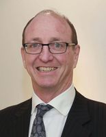
Dr James Borthwick
Ophthalmologist & Vitreo-retinal Surgeon
-

Dr Elizabeth Conner
Ophthalmologist, Corneal, Paediatric & Strabismus Specialist
-

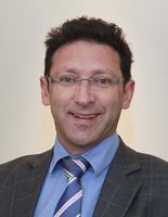
Dr Rahul Dwivedi
Ophthalmologist, Glaucoma & Cataract Specialist
-
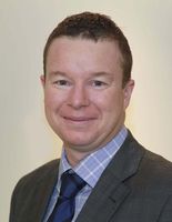
Dr Sean Every
Ophthalmologist & Vitreo-retinal Surgeon
-

Dr Genevieve Oliver
Ophthalmologist & Vitreo-retinal Surgeon
-

Dr Jo-Anne Pon
Ophthalmologist & Oculoplastic Surgeon
-
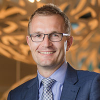
Dr John Rawstron
Ophthalmologist & Refractive Surgeon
-
Dr Logan Robinson
Ophthalmologist & Vitreo-retinal Surgeon
-

Dr Rebecca Stack
Ophthalmologist & Oculoplastic Surgeon
-
Dr Rob Weatherhead
Ophthalmologist & Oculoplastic Surgeon
-
Dr Dickson Wong
Ophthalmologist, Glaucoma, Oculoplastic and Cataract Surgeon
Referral Expectations
Appointments on referral by either General Practitioner or Optometrist are preferred but may also be made without a referral.
Please bring to your appointment:
- All glasses which you currently use for distance or near vision.
- Any letters or reports from your doctor or optometrist
- A list of all medicines you are currently taking
Fees and Charges Description
Standard Initial Consultation from $280
Hours
| Mon – Fri | 8:00 AM – 5:30 PM |
|---|
Public Holidays: Closed Labour Day (28 Oct), Canterbury Anniversary (15 Nov), Waitangi Day (6 Feb), Good Friday (18 Apr), Easter Sunday (20 Apr), Easter Monday (21 Apr), ANZAC Day (25 Apr), King's Birthday (2 Jun), Matariki (20 Jun).
Common Conditions
Cataracts are an opacity or haze in the lens of the eye. Cataract surgery replaces the cloudy lens with a clear lens. The lens becomes cloudy because the protein denatures like fresh milk becoming sour. It is a chemical change that leads to the cloudy lens and reduced eye sight. Sometimes a cataract will develop because of eye trauma, inflammation in the eye or other eye disease processes. Most often it occurs for no particular reason. A cataract that is left in the eye will slowly become more dense. It may change to a white cataract or it may in fact become brown and then black. The harder the cataract is the more energy and trauma to the eye is required to remove the cataract. The best measure of a cataract is how much it affects your eye sight. It may reduce your eye sight for fine detail such as reading or seeing the TV or it may affect your vision more markedly in bright light when driving or at night. Replacing the lens of the eye, a cataract operation, is a highly successful operation. It will allow the eye to see as well as the eye is capable of seeing in 98% of operations. If your eye has other problems such as age related macular degeneration then the cataract operation cannot restore your eye sight entirely to normal. A cataract operation does not mean you will be completely free and independent of spectacles. Spectacles fine tune the vision both for distance and near. To plan not to use spectacles is to compromise the best vision that your eye could have. A cataract operation is usually done under a local anaesthetic as a day case procedure. Southern Eye Specialists provides the postoperative eye care including medications as part of the surgical fee. This is done for the convenience of our patients. Following cataract surgery you must be seen the next day by the Eye Specialist and at three to six weeks by an eye care practitioner. When you consider having a cataract operation it is important to focus on the benefit to your eye sight at the time of the operation and in to the future against the risk of the operation. This will be discussed with you by your Eye Specialist at your first consultation appointment.
Cataracts are an opacity or haze in the lens of the eye. Cataract surgery replaces the cloudy lens with a clear lens. The lens becomes cloudy because the protein denatures like fresh milk becoming sour. It is a chemical change that leads to the cloudy lens and reduced eye sight. Sometimes a cataract will develop because of eye trauma, inflammation in the eye or other eye disease processes. Most often it occurs for no particular reason. A cataract that is left in the eye will slowly become more dense. It may change to a white cataract or it may in fact become brown and then black. The harder the cataract is the more energy and trauma to the eye is required to remove the cataract. The best measure of a cataract is how much it affects your eye sight. It may reduce your eye sight for fine detail such as reading or seeing the TV or it may affect your vision more markedly in bright light when driving or at night. Replacing the lens of the eye, a cataract operation, is a highly successful operation. It will allow the eye to see as well as the eye is capable of seeing in 98% of operations. If your eye has other problems such as age related macular degeneration then the cataract operation cannot restore your eye sight entirely to normal. A cataract operation does not mean you will be completely free and independent of spectacles. Spectacles fine tune the vision both for distance and near. To plan not to use spectacles is to compromise the best vision that your eye could have. A cataract operation is usually done under a local anaesthetic as a day case procedure. Southern Eye Specialists provides the postoperative eye care including medications as part of the surgical fee. This is done for the convenience of our patients. Following cataract surgery you must be seen the next day by the Eye Specialist and at three to six weeks by an eye care practitioner. When you consider having a cataract operation it is important to focus on the benefit to your eye sight at the time of the operation and in to the future against the risk of the operation. This will be discussed with you by your Eye Specialist at your first consultation appointment.
The best measure of a cataract is how much it affects your eye sight. It may reduce your eye sight for fine detail such as reading or seeing the TV or it may affect your vision more markedly in bright light when driving or at night.
Replacing the lens of the eye, a cataract operation, is a highly successful operation. It will allow the eye to see as well as the eye is capable of seeing in 98% of operations. If your eye has other problems such as age related macular degeneration then the cataract operation cannot restore your eye sight entirely to normal. A cataract operation does not mean you will be completely free and independent of spectacles. Spectacles fine tune the vision both for distance and near. To plan not to use spectacles is to compromise the best vision that your eye could have.
A cataract operation is usually done under a local anaesthetic as a day case procedure. Southern Eye Specialists provides the postoperative eye care including medications as part of the surgical fee. This is done for the convenience of our patients. Following cataract surgery you must be seen the next day by the Eye Specialist and at three to six weeks by an eye care practitioner.
When you consider having a cataract operation it is important to focus on the benefit to your eye sight at the time of the operation and in to the future against the risk of the operation. This will be discussed with you by your Eye Specialist at your first consultation appointment.
A strabismus, commonly known as a squint, is present when both eyes are not looking in the same direction. Sometimes, but not always, it will cause double vision. Most often it is not due to a weakness of a muscle of the eye but is due more to the messages given to the muscles by the brain. A turned eye can occur because of a wide range of causes. In children it is important that a turned eye is examined early to exclude serious disease such as an eye tumour and also to best assure that normal eye sight develops in a young child. The onset of a turned eye with or without double vision is also a serious problem in an adult as it may indicate a tumour. However there are many causes of a turned eye that are less serious. Surgical correction of a turned eye usually involves a general anaesthetic. The muscles are repositioned on the eye to correct the alignment of the two eyes. Southern Eye Specialists have extensive experience in the diagnosis and management of strabismus, double vision and the coordination of the two eyes together.
A strabismus, commonly known as a squint, is present when both eyes are not looking in the same direction. Sometimes, but not always, it will cause double vision. Most often it is not due to a weakness of a muscle of the eye but is due more to the messages given to the muscles by the brain. A turned eye can occur because of a wide range of causes. In children it is important that a turned eye is examined early to exclude serious disease such as an eye tumour and also to best assure that normal eye sight develops in a young child. The onset of a turned eye with or without double vision is also a serious problem in an adult as it may indicate a tumour. However there are many causes of a turned eye that are less serious. Surgical correction of a turned eye usually involves a general anaesthetic. The muscles are repositioned on the eye to correct the alignment of the two eyes. Southern Eye Specialists have extensive experience in the diagnosis and management of strabismus, double vision and the coordination of the two eyes together.
A strabismus, commonly known as a squint, is present when both eyes are not looking in the same direction. Sometimes, but not always, it will cause double vision.
Most often it is not due to a weakness of a muscle of the eye but is due more to the messages given to the muscles by the brain. A turned eye can occur because of a wide range of causes.
In children it is important that a turned eye is examined early to exclude serious disease such as an eye tumour and also to best assure that normal eye sight develops in a young child. The onset of a turned eye with or without double vision is also a serious problem in an adult as it may indicate a tumour.
However there are many causes of a turned eye that are less serious.
Surgical correction of a turned eye usually involves a general anaesthetic. The muscles are repositioned on the eye to correct the alignment of the two eyes.
Southern Eye Specialists have extensive experience in the diagnosis and management of strabismus, double vision and the coordination of the two eyes together.
Glaucoma is an eye disease with a characteristic damage to the optic nerve that transmits eye sight to the brain. It is the commonest cause of preventable blindness and vision loss. Multiple factors are often important in causing glaucoma, but it is most commonly related to in an increase in pressure in the eye. Symptoms are generally absent until the condition has progressed to an advanced stage. Very occasionally, a rarer form of glaucoma can develop suddenly and symptoms may then include: headaches and aches around the affected eye, seeing halos around lights, sensitivity to light, blurred vision, nausea and vomiting. You may be more likely to develop glaucoma if you: have someone else in your family with glaucoma already have high pressure in your eye have experienced injury to your eye have or have had certain other eye problems have migraine or circulation problems. Glaucoma is more common in people over 50 years of age and more common in women than men. Diagnosis usually comes after consultation with an eye doctor. Signs of glaucoma may also be picked up at an optometrist’s eye examination. The following tests are used to diagnose and monitor glaucoma: Tonometry – measures eye pressure. It is often the first screening test for glaucoma. The eyes are numbed with eye drops and then examined. Dilated eye exam - this is done with an ophthalmoscope (which is a medical instrument that allows the doctor to look through the pupil to the back of the eye).The retina and optic nerve are then examined for any sign of damage. Visual acuity test – test to check distance vision using an eye chart. Visual field test – test to measure side (peripheral) vision. Pachymetry – test to measure the thickness of the cornea. Many other new techniques are emerging to help identify the likelihood of glaucoma and help determine its rate of worsening. Although glaucoma cannot be cured, early treatment can prevent further worsening of the condition and vision loss. Regular eye examinations will need to be continued life-long. Eye drops to decrease eye pressure are the most common early treatment. Surgery may be required, especially if medications are not taking adequate effect. Laser trabeculoplasty, in which a surgeon uses a laser to help the fluid drain from the eye, may be considered in some cases, but has limited effectiveness. More commonly, a trabeculectomy may be performed when other methods have failed to adequately control pressure. This is a medium length operation that makes a new opening for fluid to drain from the eye.
Glaucoma is an eye disease with a characteristic damage to the optic nerve that transmits eye sight to the brain. It is the commonest cause of preventable blindness and vision loss. Multiple factors are often important in causing glaucoma, but it is most commonly related to in an increase in pressure in the eye. Symptoms are generally absent until the condition has progressed to an advanced stage. Very occasionally, a rarer form of glaucoma can develop suddenly and symptoms may then include: headaches and aches around the affected eye, seeing halos around lights, sensitivity to light, blurred vision, nausea and vomiting. You may be more likely to develop glaucoma if you: have someone else in your family with glaucoma already have high pressure in your eye have experienced injury to your eye have or have had certain other eye problems have migraine or circulation problems. Glaucoma is more common in people over 50 years of age and more common in women than men. Diagnosis usually comes after consultation with an eye doctor. Signs of glaucoma may also be picked up at an optometrist’s eye examination. The following tests are used to diagnose and monitor glaucoma: Tonometry – measures eye pressure. It is often the first screening test for glaucoma. The eyes are numbed with eye drops and then examined. Dilated eye exam - this is done with an ophthalmoscope (which is a medical instrument that allows the doctor to look through the pupil to the back of the eye).The retina and optic nerve are then examined for any sign of damage. Visual acuity test – test to check distance vision using an eye chart. Visual field test – test to measure side (peripheral) vision. Pachymetry – test to measure the thickness of the cornea. Many other new techniques are emerging to help identify the likelihood of glaucoma and help determine its rate of worsening. Although glaucoma cannot be cured, early treatment can prevent further worsening of the condition and vision loss. Regular eye examinations will need to be continued life-long. Eye drops to decrease eye pressure are the most common early treatment. Surgery may be required, especially if medications are not taking adequate effect. Laser trabeculoplasty, in which a surgeon uses a laser to help the fluid drain from the eye, may be considered in some cases, but has limited effectiveness. More commonly, a trabeculectomy may be performed when other methods have failed to adequately control pressure. This is a medium length operation that makes a new opening for fluid to drain from the eye.
Glaucoma is an eye disease with a characteristic damage to the optic nerve that transmits eye sight to the brain. It is the commonest cause of preventable blindness and vision loss.
Multiple factors are often important in causing glaucoma, but it is most commonly related to in an increase in pressure in the eye. Symptoms are generally absent until the condition has progressed to an advanced stage. Very occasionally, a rarer form of glaucoma can develop suddenly and symptoms may then include: headaches and aches around the affected eye, seeing halos around lights, sensitivity to light, blurred vision, nausea and vomiting.
- have someone else in your family with glaucoma
- already have high pressure in your eye
- have experienced injury to your eye
- have or have had certain other eye problems
- have migraine or circulation problems.
- Tonometry – measures eye pressure. It is often the first screening test for glaucoma. The eyes are numbed with eye drops and then examined.
- Dilated eye exam - this is done with an ophthalmoscope (which is a medical instrument that allows the doctor to look through the pupil to the back of the eye).The retina and optic nerve are then examined for any sign of damage.
- Visual acuity test – test to check distance vision using an eye chart.
- Visual field test – test to measure side (peripheral) vision.
- Pachymetry – test to measure the thickness of the cornea.
This is a complication of diabetes and is caused by small blood vessel damage within the retina of the eye. It commonly affects both eyes and may cause permanent loss of vision. Macular oedema is sometimes also present with diabetic retinopathy. Macular oedema is when fluid leaks into the retina and causes swelling and blurred vision. This may occur at any stage of diabetic retinopathy, but is more common as the disease progresses. There are often no symptoms in the early stages but as the condition progresses vision may begin to become impaired. Often visual loss may be sudden and without warning. This is why it is imperative that at-risk diabetics have frequent eye checks. Poorly controlled diabetes and pregnancy in diabetes are risk factors for developing this condition. Often, first-stage diabetic retinopathy requires no active treatment on the eye but requires stabilisation of diabetes and regular eye examinations. With progressive retinopathy, a laser treatment called the PRP laser can be used. This works by shrinking enlarged blood vessels to prevent further bleeding into the retina. Severe bleeding may require a surgical procedure called a vitrectomy, where blood is surgically removed from the eye. Treatment of macular oedema, if present, is by focal laser treatment. Vision is stabilised by reducing the degree of fluid leakage into the retina. Often more than one treatment is required.
This is a complication of diabetes and is caused by small blood vessel damage within the retina of the eye. It commonly affects both eyes and may cause permanent loss of vision. Macular oedema is sometimes also present with diabetic retinopathy. Macular oedema is when fluid leaks into the retina and causes swelling and blurred vision. This may occur at any stage of diabetic retinopathy, but is more common as the disease progresses. There are often no symptoms in the early stages but as the condition progresses vision may begin to become impaired. Often visual loss may be sudden and without warning. This is why it is imperative that at-risk diabetics have frequent eye checks. Poorly controlled diabetes and pregnancy in diabetes are risk factors for developing this condition. Often, first-stage diabetic retinopathy requires no active treatment on the eye but requires stabilisation of diabetes and regular eye examinations. With progressive retinopathy, a laser treatment called the PRP laser can be used. This works by shrinking enlarged blood vessels to prevent further bleeding into the retina. Severe bleeding may require a surgical procedure called a vitrectomy, where blood is surgically removed from the eye. Treatment of macular oedema, if present, is by focal laser treatment. Vision is stabilised by reducing the degree of fluid leakage into the retina. Often more than one treatment is required.
This is when the retina detaches, meaning it is lifted or separated from its normal position within the eye. An acute retinal detachment requires urgent assessment and appropriate treatment. Unless prompt and effective treatment is given, some forms of retinal detachment may lead to irreversible blindness. Signs and symptoms include: a sudden or gradual increase in floaters, deterioration in vision, cobwebs or specks with the visual field, light flashes in the eye or the appearance of curtains over the visual field. You are more likely to have a retinal detachment if you are very short-sighted or have had an injury or previous surgery to the eye. For minor detachments, a laser or freeze treatment (cryopexy) are used. Both therapies re-attach the retina. For major detachment, surgery will be necessary. A band is often put around the back of the eye to prevent further detachment. Surgical treatment is usually a vitrectomy, where the jelly (vitreous) is removed from the eye. This often involves a hospital stay. It can take several months post-surgery to see the final visual result.
This is when the retina detaches, meaning it is lifted or separated from its normal position within the eye. An acute retinal detachment requires urgent assessment and appropriate treatment. Unless prompt and effective treatment is given, some forms of retinal detachment may lead to irreversible blindness. Signs and symptoms include: a sudden or gradual increase in floaters, deterioration in vision, cobwebs or specks with the visual field, light flashes in the eye or the appearance of curtains over the visual field. You are more likely to have a retinal detachment if you are very short-sighted or have had an injury or previous surgery to the eye. For minor detachments, a laser or freeze treatment (cryopexy) are used. Both therapies re-attach the retina. For major detachment, surgery will be necessary. A band is often put around the back of the eye to prevent further detachment. Surgical treatment is usually a vitrectomy, where the jelly (vitreous) is removed from the eye. This often involves a hospital stay. It can take several months post-surgery to see the final visual result.
Refreshments
Complimentary Tea and Coffee available
Parking
We have on-site parking, accessible via Kilmore Street.
Pharmacy
There is a pharmacy located at Forte Hospital, across the road.
Website

Contact Details
128 Kilmore Street, Christchurch
Canterbury
-
Phone
(03) 355 6397
-
Fax
(03) 355 6156
Healthlink EDI
Southeye
Email
Website
Use our online contact form
Hours: Monday to Friday 8.15am to 5.15pm
128 Kilmore Street
Christchurch Central
Christchurch
Canterbury 8013
Street Address
128 Kilmore Street
Christchurch Central
Christchurch
Canterbury 8013
Postal Address
PO Box 21023
Christchurch 8140
Was this page helpful?
This page was last updated at 10:54AM on July 16, 2024. This information is reviewed and edited by Southern Eye Specialists.