Canterbury > Private Hospitals & Specialists >
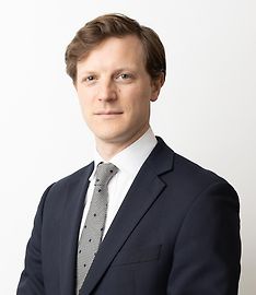
Andrew Powell - Orthopaedic Surgeon
Private Service, Orthopaedics
Today
Description
What is Orthopaedics?
Staff
Practice manager: Emma-Lee Davidson
Consultants
-

Mr Andrew Powell
Orthopaedic Surgeon
How do I access this service?
Referral
Mr Powell typically receives GP referrals, but direct patient enquiries are welcome.
Contact us
Contact our Practice Manager, Emma-Lee Davidson
Ph: 022 192 6078
Email: admin@ajpowellorthopaedics.com
Referral Expectations
You need to bring with you:
Fees and Charges Categorisation
Fees apply
Fees and Charges Description
Mr Powell is a Southern Cross Affiliated Provider, and NIB First Choice member.
Hours
| Mon – Fri | 8:30 AM – 5:30 PM |
|---|
Reception opening hours are 8:30am till 5:30pm
Procedures / Treatments
Orthopaedic surgeons have expertise in the treatment of fractured (broken) bones, particularly in the assessment of damage that may have occurred around the fracture. Follow-up of a fracture may involve monitoring the progress of the healing bone, checking the position of the bone in a cast and deciding when other steps in management such as re-manipulation of the fracture or removal of a cast is required. Click here for more information about fractures.
Orthopaedic surgeons have expertise in the treatment of fractured (broken) bones, particularly in the assessment of damage that may have occurred around the fracture. Follow-up of a fracture may involve monitoring the progress of the healing bone, checking the position of the bone in a cast and deciding when other steps in management such as re-manipulation of the fracture or removal of a cast is required. Click here for more information about fractures.
Orthopaedic surgeons have expertise in the treatment of fractured (broken) bones, particularly in the assessment of damage that may have occurred around the fracture.
Follow-up of a fracture may involve monitoring the progress of the healing bone, checking the position of the bone in a cast and deciding when other steps in management such as re-manipulation of the fracture or removal of a cast is required.
Click here for more information about fractures.
For elderly patients joint replacement surgery is commonly required to treat damaged joints from wearing out, arthritis or other forms of joint disease including rheumatoid arthritis. In these procedures the damaged joint surface is removed and replaced with artificial surfaces normally made from metal (chromium cobalt alloy, titanium), plastic (high density polyethelene) or ceramic which act as alternate bearing surfaces for the damaged joint. These operations are major procedures which require the patient to be in hospital for several days and followed by a significant period of rehabilitation. The hospital has several ways of approaching the procedure for replacement and the specifics for the procedure will be covered at the time of assessment and booking of surgery. Occasionally blood transfusions are required; if you have some concerns raise this with your surgeon during consultation.
For elderly patients joint replacement surgery is commonly required to treat damaged joints from wearing out, arthritis or other forms of joint disease including rheumatoid arthritis. In these procedures the damaged joint surface is removed and replaced with artificial surfaces normally made from metal (chromium cobalt alloy, titanium), plastic (high density polyethelene) or ceramic which act as alternate bearing surfaces for the damaged joint. These operations are major procedures which require the patient to be in hospital for several days and followed by a significant period of rehabilitation. The hospital has several ways of approaching the procedure for replacement and the specifics for the procedure will be covered at the time of assessment and booking of surgery. Occasionally blood transfusions are required; if you have some concerns raise this with your surgeon during consultation.
This is a surgical procedure performed on a knee joint that has become painful and/or impaired because of disease, injury or wear and tear. In total knee replacement, artificial materials (metal and plastic) are used to replace the following damaged surfaces within the knee joint: the end of the thigh bone (femur) the end of the shin bone (tibia) the back of the kneecap (patella) This operation is a major procedure which requires you to be in hospital for several days and will be followed by a significant period of rehabilitation. Occasionally blood transfusions are required; if you have some concerns raise this with your surgeon during consultation. For more information about total knee replacement please click here.
This is a surgical procedure performed on a knee joint that has become painful and/or impaired because of disease, injury or wear and tear. In total knee replacement, artificial materials (metal and plastic) are used to replace the following damaged surfaces within the knee joint: the end of the thigh bone (femur) the end of the shin bone (tibia) the back of the kneecap (patella) This operation is a major procedure which requires you to be in hospital for several days and will be followed by a significant period of rehabilitation. Occasionally blood transfusions are required; if you have some concerns raise this with your surgeon during consultation. For more information about total knee replacement please click here.
This is a surgical procedure performed on a knee joint that has become painful and/or impaired because of disease, injury or wear and tear.
In total knee replacement, artificial materials (metal and plastic) are used to replace the following damaged surfaces within the knee joint:
- the end of the thigh bone (femur)
- the end of the shin bone (tibia)
- the back of the kneecap (patella)
This operation is a major procedure which requires you to be in hospital for several days and will be followed by a significant period of rehabilitation.
Occasionally blood transfusions are required; if you have some concerns raise this with your surgeon during consultation.
For more information about total knee replacement please click here.
The anterior cruciate ligament (ACL) is a strong, stabilising ligament running through the centre of the knee between the femur (thigh bone) and tibia (shin bone). When the ACL is torn, frequently as the result of a sporting injury, arthroscopic surgery known as ACL Reconstruction is performed. The procedure involves replacement of the damaged ligament with tissue grafted from elsewhere, usually the patellar or hamstring tendon. The ends of the grafted tendon are attached to the femur at one end and the tibia at the other using screws or staples. For more information about ACL Reconstruction please click here.
The anterior cruciate ligament (ACL) is a strong, stabilising ligament running through the centre of the knee between the femur (thigh bone) and tibia (shin bone). When the ACL is torn, frequently as the result of a sporting injury, arthroscopic surgery known as ACL Reconstruction is performed. The procedure involves replacement of the damaged ligament with tissue grafted from elsewhere, usually the patellar or hamstring tendon. The ends of the grafted tendon are attached to the femur at one end and the tibia at the other using screws or staples. For more information about ACL Reconstruction please click here.
The anterior cruciate ligament (ACL) is a strong, stabilising ligament running through the centre of the knee between the femur (thigh bone) and tibia (shin bone).
When the ACL is torn, frequently as the result of a sporting injury, arthroscopic surgery known as ACL Reconstruction is performed. The procedure involves replacement of the damaged ligament with tissue grafted from elsewhere, usually the patellar or hamstring tendon. The ends of the grafted tendon are attached to the femur at one end and the tibia at the other using screws or staples.
For more information about ACL Reconstruction please click here.
This procedure is used when osteoarthritic damage to the cartilage on one side of the knee has caused the angle of the knee joint to change so that most of the body's weight is borne by the affected side, adding to the wear on that side. High Tibial Osteotomy involves reshaping and realignment of the bone so that weight becomes more evenly distributed between the inside and outside of the knee, thereby reducing the workload on the damaged side. You will probably have to stay in hospital for several days after surgery followed by up to 6 months rehabilitation. For more information about osteotomy please click here.
This procedure is used when osteoarthritic damage to the cartilage on one side of the knee has caused the angle of the knee joint to change so that most of the body's weight is borne by the affected side, adding to the wear on that side. High Tibial Osteotomy involves reshaping and realignment of the bone so that weight becomes more evenly distributed between the inside and outside of the knee, thereby reducing the workload on the damaged side. You will probably have to stay in hospital for several days after surgery followed by up to 6 months rehabilitation. For more information about osteotomy please click here.
This procedure is used when osteoarthritic damage to the cartilage on one side of the knee has caused the angle of the knee joint to change so that most of the body's weight is borne by the affected side, adding to the wear on that side.
High Tibial Osteotomy involves reshaping and realignment of the bone so that weight becomes more evenly distributed between the inside and outside of the knee, thereby reducing the workload on the damaged side.
You will probably have to stay in hospital for several days after surgery followed by up to 6 months rehabilitation.
For more information about osteotomy please click here.
The menisci are two circular strips of cartilage that form a cushioning layer between the ends of the femur (thigh bone) and tibia (shin bone) in the knee joint. Together the medial and lateral menisci, on the inside and outside of the knee, respectively, act as shock absorbers and distribute the weight of the body across the knee joint. The menisci can become torn through injury or damaged from age-related wear and tear and may require surgery. The most common meniscal surgery is partial meniscectomy in which the torn portion of the meniscus is cut away so that the cartilage surface is smooth again. In some cases meniscal repair is carried out, in this case the torn edges of the meniscus are sutured together. Both procedures are performed arthroscopically. For more information please click here for meniscal tears and click here for meniscal transplant surgery.
The menisci are two circular strips of cartilage that form a cushioning layer between the ends of the femur (thigh bone) and tibia (shin bone) in the knee joint. Together the medial and lateral menisci, on the inside and outside of the knee, respectively, act as shock absorbers and distribute the weight of the body across the knee joint. The menisci can become torn through injury or damaged from age-related wear and tear and may require surgery. The most common meniscal surgery is partial meniscectomy in which the torn portion of the meniscus is cut away so that the cartilage surface is smooth again. In some cases meniscal repair is carried out, in this case the torn edges of the meniscus are sutured together. Both procedures are performed arthroscopically. For more information please click here for meniscal tears and click here for meniscal transplant surgery.
The menisci are two circular strips of cartilage that form a cushioning layer between the ends of the femur (thigh bone) and tibia (shin bone) in the knee joint. Together the medial and lateral menisci, on the inside and outside of the knee, respectively, act as shock absorbers and distribute the weight of the body across the knee joint.
The menisci can become torn through injury or damaged from age-related wear and tear and may require surgery.
The most common meniscal surgery is partial meniscectomy in which the torn portion of the meniscus is cut away so that the cartilage surface is smooth again.
In some cases meniscal repair is carried out, in this case the torn edges of the meniscus are sutured together.
Both procedures are performed arthroscopically.
For more information please click here for meniscal tears and click here for meniscal transplant surgery.
Over the last 30 years a large number of orthopaedic procedures on joints have been performed using an arthroscope, where a fiber optic telescope is used to look inside the joint. Through this type of keyhole surgery, fine instruments can be introduced through small incisions (portals) to allow surgery to be performed without the need for large cuts. This allows many procedures to be performed as a day stay and allows quicker return to normal function of the joint. Arthroscopic surgery is less painful than open surgery and decreases the risk of healing problems. Arthroscopy allows access to parts of the joints which can not be accessed by other types of surgery.
Over the last 30 years a large number of orthopaedic procedures on joints have been performed using an arthroscope, where a fiber optic telescope is used to look inside the joint. Through this type of keyhole surgery, fine instruments can be introduced through small incisions (portals) to allow surgery to be performed without the need for large cuts. This allows many procedures to be performed as a day stay and allows quicker return to normal function of the joint. Arthroscopic surgery is less painful than open surgery and decreases the risk of healing problems. Arthroscopy allows access to parts of the joints which can not be accessed by other types of surgery.
In many cases tendons will be lengthened to improve the muscle balance around a joint or tendons will be transferred to give overall better joint function. This occurs in children with neuromuscular conditions but also applies to a number of other conditions. Most of these procedures involve some sort of splintage after the surgery followed by a period of rehabilitation, normally supervised by a physiotherapist.
In many cases tendons will be lengthened to improve the muscle balance around a joint or tendons will be transferred to give overall better joint function. This occurs in children with neuromuscular conditions but also applies to a number of other conditions. Most of these procedures involve some sort of splintage after the surgery followed by a period of rehabilitation, normally supervised by a physiotherapist.
Orthopaedic deformities can be congenital or acquired as the result of injury, infection or tumour. Resulting in crooked limbs or discrepancies in limb length, such deformities can affect appearance and function and can often cause significant pain. Osteotomy is the division of a crooked or bent bone to improve alignment of the limb. These procedures normally involve some form of internal fixation, such as rods or plates, or external fixation which involves external wires and pins to hold the bone. The type of procedure for fixation will be explained when the surgery is planned. Some of the more common orthopaedic deformities are: Intoeing Bow Legs (Genu Varum) Club Foot (Talipes) Developmental Dislocation of the Hip Bunions Limb Length Discrepancy
Orthopaedic deformities can be congenital or acquired as the result of injury, infection or tumour. Resulting in crooked limbs or discrepancies in limb length, such deformities can affect appearance and function and can often cause significant pain. Osteotomy is the division of a crooked or bent bone to improve alignment of the limb. These procedures normally involve some form of internal fixation, such as rods or plates, or external fixation which involves external wires and pins to hold the bone. The type of procedure for fixation will be explained when the surgery is planned. Some of the more common orthopaedic deformities are: Intoeing Bow Legs (Genu Varum) Club Foot (Talipes) Developmental Dislocation of the Hip Bunions Limb Length Discrepancy
Orthopaedic deformities can be congenital or acquired as the result of injury, infection or tumour. Resulting in crooked limbs or discrepancies in limb length, such deformities can affect appearance and function and can often cause significant pain.
Osteotomy is the division of a crooked or bent bone to improve alignment of the limb. These procedures normally involve some form of internal fixation, such as rods or plates, or external fixation which involves external wires and pins to hold the bone. The type of procedure for fixation will be explained when the surgery is planned.
Some of the more common orthopaedic deformities are:
Disability Assistance
Wheelchair access, Wheelchair accessible toilet, Mobility parking space
Parking
Free street parking nearby, or paid parking at the hospital
Pharmacy

Contact Details
165 Leinster Rd, Christchurch
Canterbury
-
Phone
(022) 192 6078
Healthlink EDI
AJPOWELL
Email
Practice manager: Emma-Lee Davidson
Leinster Orthopaedics, St George's Hospital, 165 Leinster Road
Merivale
Christchurch
Canterbury 8146
Street Address
Leinster Orthopaedics, St George's Hospital, 165 Leinster Road
Merivale
Christchurch
Canterbury 8146
Postal Address
Leinster Orthopaedics
St George’s Hospital
165 Leinster Road
Christchurch 8014
Was this page helpful?
This page was last updated at 3:23PM on November 21, 2023. This information is reviewed and edited by Andrew Powell - Orthopaedic Surgeon.
